
Why Did My Doctor Order a Calcium Test Along With Vitamin D?
Calcium and vitamin D show up together on so many lab orders that it's easy to assume they're just routinely bundled the way a basic metabolic panel bundles a dozen unrelated values. They're not. Vitamin D's single most important job in your entire body is making calcium absorption from your gut possible in the first place — without enough of it, your intestines simply can't pull enough calcium out of the food you eat, no matter how much calcium that food actually contains. Because of this direct, one-way dependency, a calcium result on its own and a vitamin D result on its own each tell only half of a story that only makes complete sense when you read them together. This article walks through the actual absorption chain connecting these two values, the four genuinely distinct patterns a doctor looks for when reading them side by side, and the specific situations — from malabsorption to bone density monitoring to vitamin D toxicity — where testing them together, rather than separately, actually changes what happens next.
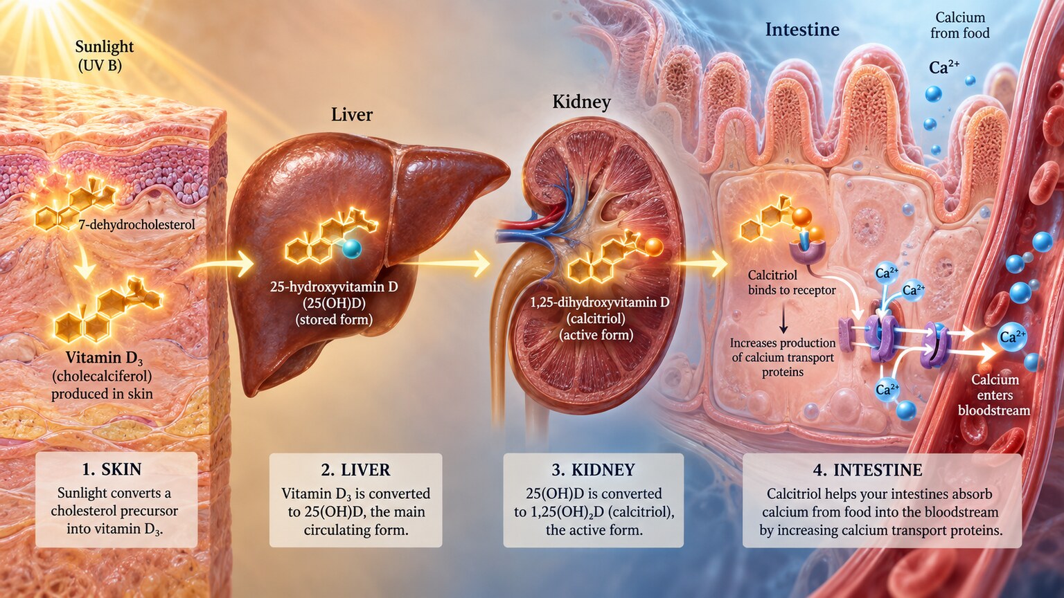
Figure 1. Vitamin D activated by the liver and kidneys instructs intestinal cells to build the specific transport proteins needed to actually pull calcium out of digested food and into the bloodstream.
Already have a calcium number from a recent test, but not sure if it's actually good or bad for you? Just plug it in and see instantly.
🧮 Try the Free CalculatorThe One-Way Dependency: Why Calcium Absorption Needs Vitamin D at All
Calcium doesn't cross the lining of your small intestine on its own, no matter how much of it sits in your gut after a meal. The cells lining your intestine need to actively build specific transport proteins — most notably one called calbindin — that grab calcium ions from digested food and carry them across the intestinal wall into your bloodstream. Building these transport proteins is a job assigned specifically to activated vitamin D, which binds to a receptor inside intestinal cells and switches on the genes responsible for producing them. Without enough activated vitamin D circulating, this genetic switch stays largely off, and the intestine's capacity to absorb calcium drops dramatically — research suggests calcium absorption efficiency can fall from around 30-40% of dietary intake in a person with adequate vitamin D down to somewhere in the range of 10-15% in someone who's significantly deficient, even when their diet contains exactly the same amount of calcium either way.
This is the core reason these two tests are so often ordered as a pair rather than independently: a low calcium result can't be fully interpreted without knowing whether vitamin D deficiency is the underlying reason absorption has been compromised, and a low vitamin D result can't be fully evaluated for its downstream consequences without checking whether calcium absorption has actually been affected yet. They're measuring two different points along the exact same physiological pathway, and a problem at the vitamin D end of that pathway doesn't necessarily show up as a problem at the calcium end immediately — which is precisely what the next section explains.
Why Vitamin D Has to Be Activated Twice Before It Can Do This Job
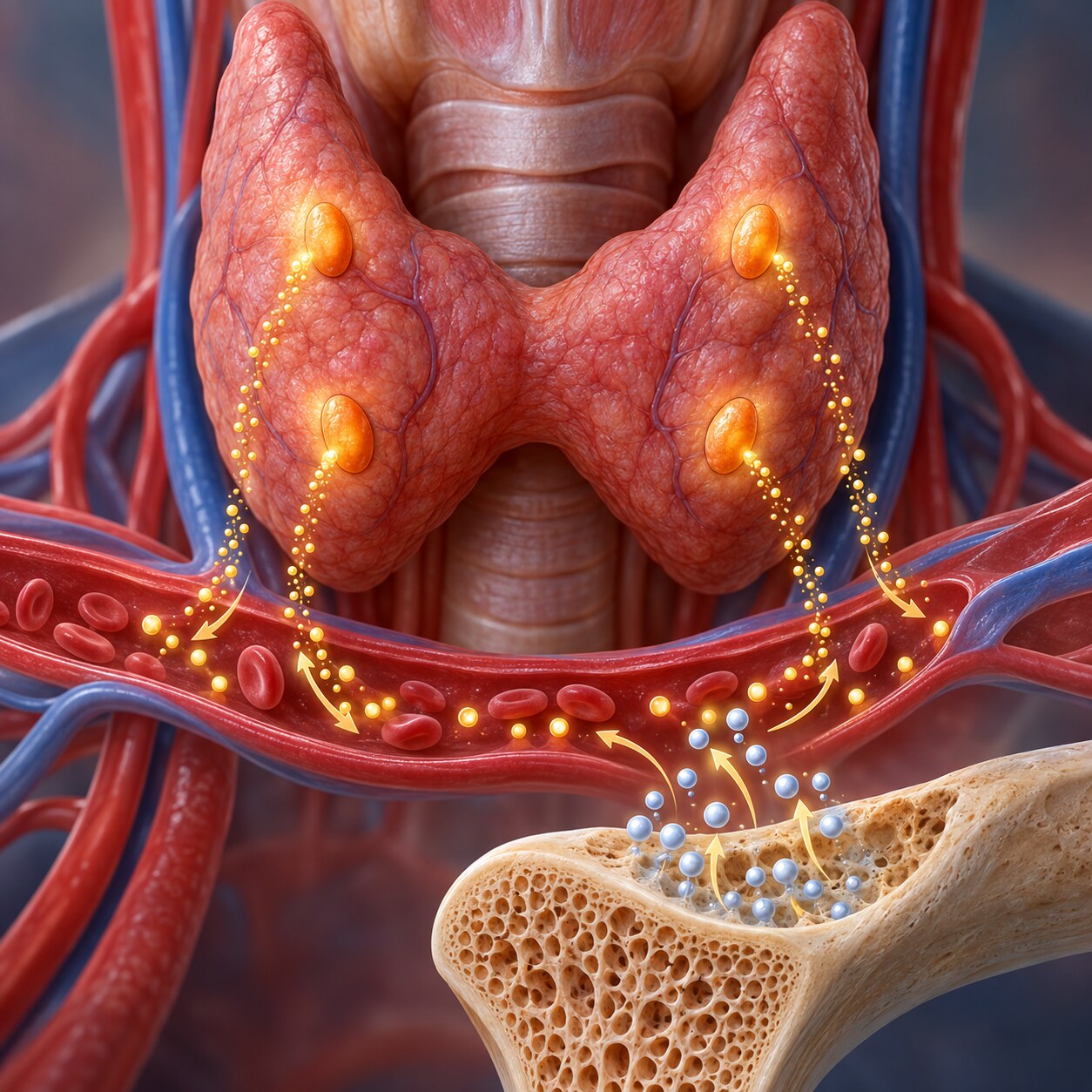
Figure 2. Four rice-grain-sized parathyroid glands continuously monitor blood calcium and release parathyroid hormone whenever levels drift downward, forming a tight feedback loop with vitamin D.
The vitamin D your skin makes from sun exposure, or that you get from food and supplements, isn't biologically active in the form it first enters your bloodstream. It has to pass through two separate conversion steps before it can switch on calcium absorption in the gut. The first conversion happens in the liver, which turns raw vitamin D into 25-hydroxyvitamin D — the specific, stable form that a standard vitamin D blood test actually measures, precisely because it's the most reliable indicator of your overall vitamin D stores. The second, final conversion happens in the kidneys, which turn that liver-processed form into the fully activated hormone, calcitriol, that actually does the work of switching on intestinal calcium absorption.
This second kidney conversion step is tightly controlled by parathyroid hormone, released by four tiny glands sitting just behind your thyroid, which continuously monitor blood calcium levels and ramp up this final activation step specifically when calcium is running low. This feedback relationship is exactly why a person can have a genuinely low vitamin D storage level (the 25-hydroxyvitamin D your test measures) while their calcium still reads normal for a while: parathyroid hormone compensates by squeezing more activation out of whatever vitamin D is available, plus pulling additional calcium out of bone reserves, propping up blood calcium even as the underlying vitamin D deficiency continues. This compensation is genuinely useful in the short term, but it isn't free — the calcium being pulled out of bone to maintain that normal blood level is calcium your skeleton needed to keep, which is a large part of why long-standing vitamin D deficiency is so strongly linked to bone loss even in people whose calcium blood tests have looked normal the entire time.
The Four Patterns: What Reading Both Results Together Actually Reveals
Because of this absorption chain and the compensation described above, looking at calcium and vitamin D together reveals one of four genuinely distinct clinical pictures, each of which points toward a different explanation and a different next step.
Pattern one: normal vitamin D, normal calcium. This is the reassuring baseline — the absorption chain is working as expected, and no further evaluation related to this specific pathway is generally needed.
Pattern two: low vitamin D, low calcium. This is the pattern that most directly confirms the mechanism this article has described: vitamin D deficiency has progressed far enough, or developed quickly enough, that the parathyroid compensation described above hasn't been able to fully keep blood calcium propped up. This combination generally points toward correcting the vitamin D deficiency directly, often alongside calcium supplementation in more significant cases, since simply raising vitamin D alone can take time to restore absorption fully.
Pattern three: low vitamin D, normal calcium. This is actually the most common pattern seen in otherwise healthy people with vitamin D deficiency, and it reflects the compensation mechanism working exactly as designed — parathyroid hormone is quietly picking up the slack. A normal calcium result in this pattern is not a reason to dismiss the low vitamin D as unimportant; it's a sign the compensation is still holding, not that no problem exists. Left uncorrected, this pattern is part of why chronic vitamin D deficiency is linked to gradually declining bone density over years, even in people who never see an abnormal calcium result along the way.
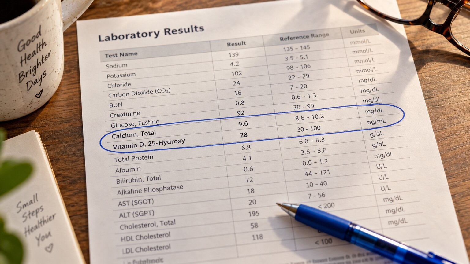
Figure 3. Reading calcium and vitamin D as a pair, rather than scanning each row in isolation, is what actually reveals which of the four underlying patterns a given result represents.
Pattern four: high calcium, checked against vitamin D. This pattern runs in the opposite direction from the other three, and it's specifically why an unexpectedly high calcium result often prompts a vitamin D check rather than the reverse. Taking extremely large doses of vitamin D supplements over time — well beyond typical recommended amounts, most often from megadose over-the-counter products taken without medical supervision — can push vitamin D activation high enough to drive excessive calcium absorption, leading to genuine vitamin D toxicity and dangerously elevated blood calcium. Checking vitamin D alongside an unexplained high calcium result is one of the more important, sometimes overlooked reasons for ordering the pair together, since this specific cause is both entirely avoidable and easy to miss if vitamin D isn't checked at the same time.
Vitamin D Toxicity: An Avoidable Cause of Dangerously High Calcium

Figure 4. Vitamin D toxicity almost always traces back to sustained, unsupervised megadose supplementation rather than diet or sun exposure, since the body has a natural ceiling on how much vitamin D it produces from sunlight alone.
It's worth understanding specifically why vitamin D toxicity is a supplement problem and virtually never a sun-exposure or dietary problem. Your skin has a built-in safety mechanism: prolonged sun exposure actually degrades excess vitamin D precursor into inactive byproducts once enough has already been produced, meaning it's essentially impossible to overdose on vitamin D from sunlight alone, no matter how long you stay outside. Diet alone rarely supplies enough vitamin D to cause toxicity either, since even vitamin-D-fortified foods contain relatively modest amounts. Toxicity is almost exclusively a supplement phenomenon, typically requiring sustained daily doses many times higher than standard recommended amounts, taken for weeks to months, often from a manufacturing error, a misunderstanding of dosing units, or someone self-treating suspected deficiency with increasingly large doses without follow-up testing to check whether the dose is actually appropriate.
Once vitamin D toxicity develops, the resulting hypercalcemia can produce genuinely serious symptoms — nausea, vomiting, excessive thirst and urination, confusion, and in severe, prolonged cases, kidney damage from calcium deposits forming inside kidney tissue itself. Recognizing this specific cause matters because the treatment is straightforward once identified: stopping the supplement and allowing vitamin D levels to gradually decline, sometimes alongside more active treatment for significantly elevated calcium in severe cases, resolves the problem in a way that wouldn't happen if the elevated calcium were instead mistakenly attributed to a completely different cause, like primary hyperparathyroidism, which requires an entirely different treatment approach.
Conditions That Disrupt This Pathway From the Absorption Side
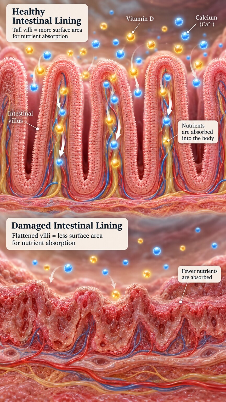
Figure 5. Conditions that damage or bypass the small intestine's absorptive lining can block calcium uptake even when vitamin D activation itself is working normally, requiring a different diagnostic approach than simple deficiency.
Because both vitamin D and calcium ultimately have to be absorbed through the same stretch of small intestine, several conditions that damage or bypass that absorptive surface disrupt this entire pathway regardless of how much vitamin D is actually present in the bloodstream. Celiac disease, an autoimmune reaction to gluten, damages the small, finger-like projections called villi that line the intestine and dramatically increase its absorptive surface area — when these villi flatten out from ongoing inflammation, both calcium and fat-soluble vitamin D absorption drop significantly, which is exactly why calcium and vitamin D deficiency are both common, and often the first clue, in someone with undiagnosed celiac disease. Bariatric surgery, particularly procedures that reroute or bypass sections of the small intestine, produces a similar effect through mechanical rather than inflammatory means, which is why patients who've had these procedures require lifelong monitoring of both values together rather than either one alone. Chronic pancreatic insufficiency and inflammatory bowel disease affecting the small intestine can produce a comparable pattern through their own distinct mechanisms.
In any of these malabsorption scenarios, simply increasing vitamin D and calcium intake through diet or standard-dose supplements often isn't enough, since the underlying absorptive surface itself is compromised — treatment typically requires either much higher supplemental doses, specific forms of vitamin D or calcium that bypass some of the affected absorption steps, or, most importantly, treating the underlying condition itself (a gluten-free diet for celiac disease, for example) to allow the intestinal lining to heal and absorption to genuinely recover over time.
Bone Density Monitoring: Where This Pairing Becomes an Ongoing Routine
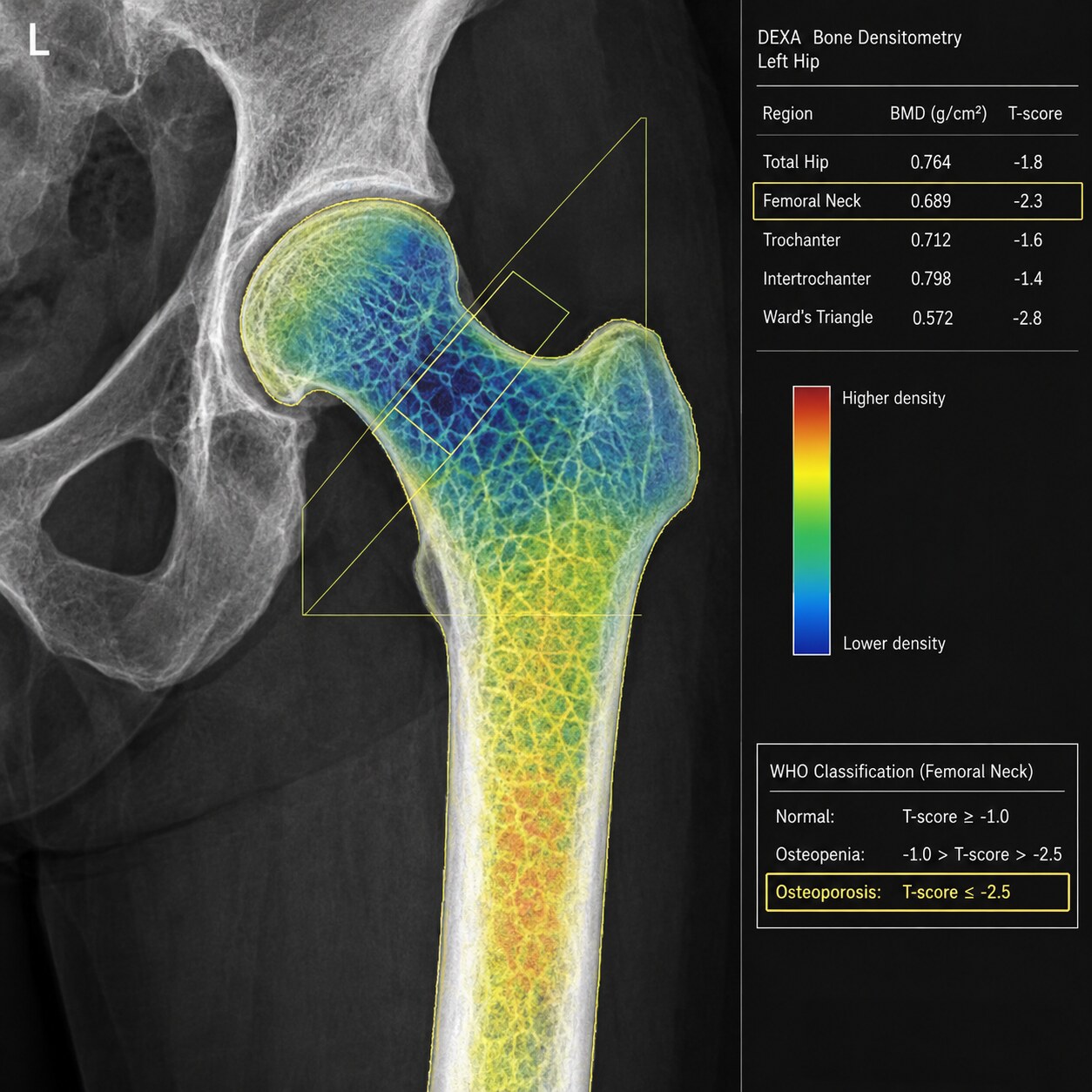
Figure 6. Confirming that calcium and vitamin D are both adequate before and during osteoporosis treatment ensures that bone-building medications actually have the raw materials they need to work.
Beyond diagnosing an acute problem, calcium and vitamin D are checked together routinely as part of monitoring bone health, particularly in anyone diagnosed with osteoporosis or low bone density on a bone density scan. This pairing matters here for a very practical reason: the medications used to treat osteoporosis, most commonly a class called bisphosphonates, work by slowing the breakdown of existing bone, but they don't supply any new calcium or vitamin D themselves. Starting one of these medications while a person is still calcium- or vitamin-D-deficient can, in some cases, actually worsen low blood calcium, since the medication is simultaneously slowing bone's ability to release its own calcium reserves as a backup source. This is exactly why guidelines recommend confirming adequate calcium and vitamin D status before starting bone-density medication, and continuing to monitor both periodically throughout treatment, rather than assuming the medication alone is doing everything necessary to protect bone health.
How This Differs From a Routine Vitamin D Check for Deficiency Symptoms
It's worth distinguishing this specific pairing from a vitamin D test ordered on its own for general symptoms like fatigue or during a routine wellness visit, where calcium may not be checked at the same time simply because there's no specific reason yet to suspect the absorption chain has been affected. Calcium gets added to the order specifically when there's a reason to think the downstream effect of vitamin D status might already be showing up — existing bone density concerns, symptoms suggestive of calcium imbalance like muscle cramping or unusual fatigue beyond what vitamin D deficiency alone typically causes, a known malabsorption condition, osteoporosis treatment planning, or an unexplained abnormal calcium result that needs vitamin D checked to help explain it. Understanding this distinction helps explain why one person's vitamin D test comes back as a single number while another person's comes back paired with calcium on the same report — it generally reflects a different starting clinical question, not an inconsistency in how the test is ordered.
Who Actually Ends Up With This Combination Ordered
Certain groups of people are considerably more likely to have calcium and vitamin D checked together as a routine matter, rather than only after a problem is already suspected, and understanding who falls into these groups helps explain why the pairing shows up so often in everyday clinical practice. Older adults are checked together routinely because both aging skin's reduced capacity to synthesize vitamin D from sunlight and age-related declines in kidney function (which handles that final activation step) compound to make this population disproportionately likely to land in one of the deficiency patterns described earlier. Pregnant and breastfeeding women are another group checked together proactively, since fetal and infant bone development places substantial additional demand on the mother's calcium and vitamin D reserves, and a deficiency that might be mild and unnoticed in a non-pregnant adult can meaningfully affect both maternal bone health and infant development if it goes uncorrected.
People with darker skin pigmentation are also more likely to have this pairing checked proactively, since melanin — the same pigment that provides protection from UV damage — also reduces the skin's efficiency at synthesizing vitamin D from a given amount of sun exposure, meaning equivalent sun exposure produces less vitamin D synthesis than it would in someone with lighter skin. This is a well-documented population-level pattern, though it's worth noting it operates on a completely separate mechanism from the absorption chain covered throughout most of this article — it affects how much vitamin D gets made in the first place, not how effectively existing vitamin D drives calcium absorption once it's already circulating. Anyone taking long-term corticosteroid medications for a chronic inflammatory condition falls into this proactively-monitored group as well, since corticosteroids are well known to interfere with calcium absorption and accelerate bone loss through mechanisms partly independent of vitamin D status, making the combined picture especially important to track in this population.
A Practical Walkthrough: Two Worked Examples
Seeing these patterns applied to specific, realistic numbers makes the framework considerably easier to apply to an actual lab report. Consider someone whose report shows a 25-hydroxyvitamin D of 14 ng/mL (clearly deficient, since most labs flag anything under roughly 20 ng/mL as deficient) alongside a total calcium of 8.2 mg/dL (below a typical reference range of about 8.5 to 10.5 mg/dL). This combination fits pattern two described earlier: deficiency severe enough that parathyroid compensation hasn't been able to fully maintain normal blood calcium. This person would typically be started on vitamin D repletion, often alongside calcium supplementation given how low the calcium itself has already dropped, with follow-up testing scheduled to confirm both values are trending back into normal range together.
Now consider a second person whose report shows the same vitamin D level, 14 ng/mL, but a calcium of 9.4 mg/dL — comfortably normal. This is pattern three: the same underlying vitamin D deficiency, but with parathyroid hormone successfully compensating so far. This person still needs their vitamin D deficiency treated, and arguably benefits from catching it at this earlier stage before it has fully overwhelmed the body's compensation and started drawing down bone reserves more aggressively — but a clinician glancing only at the normal calcium result, without the vitamin D result sitting right alongside it, could easily and mistakenly conclude nothing here needs attention at all. This comparison is exactly why the framework in this article emphasizes reading the pair together rather than scanning each number against its own reference range in isolation.
What About Calcium and Vitamin D From Supplements Interacting With the Test Itself
A practical question worth addressing directly: does taking a calcium or vitamin D supplement shortly before a blood draw skew the result enough to matter? For vitamin D, the answer is generally no in any meaningful way — 25-hydroxyvitamin D reflects your body's stored levels built up over roughly the preceding month or two, not what you took that morning, since it takes time for a single dose to be processed through the liver and accumulate into the circulating pool the test actually measures. Calcium behaves somewhat differently: a very large, recent calcium supplement dose can theoretically cause a brief, small uptick in blood calcium, though in practice this effect is generally too small and too short-lived to meaningfully change a standard total calcium result taken at a routine, non-fasting blood draw. Because of this, most labs don't require stopping either supplement before testing, though it's still reasonable and often recommended practice to mention current supplement use and dosing to whoever is ordering or interpreting the test, particularly when a result comes back unexpectedly high and vitamin D megadosing needs to be considered as a possible explanation, exactly as described earlier in this article.
What This Means for Reading Your Own Combined Results
If you're looking at your own lab report and see both calcium and vitamin D listed, the practical approach is to read them together using the four-pattern framework covered earlier rather than checking each one in isolation against its own reference range. A low vitamin D with a normal calcium is not a "pass" that makes the vitamin D result unimportant — it likely reflects successful short-term compensation that's worth correcting before it progresses further. A low vitamin D with a low calcium reinforces that the deficiency has been significant enough, or gone on long enough, to overwhelm that compensation. And a high calcium result should prompt genuine curiosity about your vitamin D intake, particularly if you've been taking high-dose supplements without recent follow-up testing, before assuming a more complicated explanation is required.
It's also worth building a simple habit around any future testing: whenever one of these two values is checked, ask whether the other was included on the same order, and if not, whether there's a specific reason it wasn't. In many everyday, low-risk situations there's a perfectly good reason only one was needed — but in anyone with existing bone health concerns, a malabsorption condition, high-dose supplement use, or an unexplained abnormal result on either side, requesting the paired test rather than settling for half the picture is a reasonable, informed thing to bring up directly with whoever is managing your care.
None of this replaces an actual conversation with a healthcare provider, but knowing which of these four patterns your own results fall into gives you a genuinely useful starting point for that conversation, rather than trying to make sense of two numbers that only tell their full story when read as a pair.
How This Pathway Was Actually Discovered
The link between vitamin D and calcium absorption wasn't understood as a single connected pathway until well into the twentieth century, and the history behind it explains why these two tests are so deeply intertwined in modern clinical practice rather than being an arbitrary pairing convention. Rickets — a childhood bone-softening disease caused by severe vitamin D deficiency — was recognized clinically for centuries before its cause was understood, often misattributed to poor general nutrition or lack of exercise rather than a specific, identifiable deficiency. It wasn't until the early 1900s that researchers definitively connected rickets to both diet and sunlight exposure, and it took several more decades of biochemistry research through the mid-twentieth century to work out the specific two-step liver-then-kidney activation process this article describes, along with vitamin D's precise role in switching on intestinal calcium transport proteins at the molecular level.
This gradual scientific unraveling is part of why vitamin D testing itself is a comparatively recent addition to routine lab panels compared to calcium, which has been measurable and clinically monitored for considerably longer. Once the full mechanistic picture connecting vitamin D activation to calcium absorption was established with real biochemical detail, ordering the two together stopped being an assumption based on general nutritional overlap and became a deliberate clinical practice grounded in a specific, well-characterized physiological pathway — the same pathway this article has walked through from sunlight and diet all the way to the transport proteins inside intestinal cells.
How Kidney Function Adds a Second Layer to This Same Pathway
Because the final activation step converting stored vitamin D into its fully active hormone form happens specifically in the kidneys, chronic kidney disease disrupts this pathway through a mechanism distinct from both simple deficiency and malabsorption. In someone with significantly reduced kidney function, the kidneys may simply lack the capacity to perform this final activation step efficiently, regardless of how much stored 25-hydroxyvitamin D is actually present in the bloodstream — meaning a person can have an entirely adequate vitamin D storage level on paper while still suffering from a functional shortage of the activated hormone that actually drives calcium absorption. This is exactly why calcium and vitamin D are monitored particularly closely, often alongside phosphate and parathyroid hormone as well, in anyone with moderate to advanced chronic kidney disease, and why some patients in this situation are prescribed the already-activated form of vitamin D directly (calcitriol) rather than the standard supplement form, specifically to bypass a kidney conversion step that may no longer be working reliably.
This kidney-specific complication also explains why chronic kidney disease is one of the more complicated settings for interpreting calcium and vitamin D together, since multiple parts of the same regulatory system can be disrupted simultaneously — reduced activation capacity, altered phosphate handling, and blunted parathyroid hormone responsiveness can all be happening at once, in ways that don't fit neatly into the four simpler patterns described earlier in this article for someone with otherwise normal kidney function. Nephrologists managing this population typically follow more specialized, kidney-disease-specific guidelines for interpreting and treating abnormal results in this combination, precisely because the standard framework doesn't fully capture everything going on physiologically once kidney function itself becomes part of the equation.
Age-Related Reference Range Differences Worth Knowing
Both calcium and vitamin D reference ranges shift somewhat across the lifespan, and understanding these shifts helps explain why the same raw number can be interpreted differently depending on a person's age. Children and adolescents undergoing active bone growth generally run slightly higher calcium levels than adults, reflecting the elevated bone turnover happening during this growth period, and vitamin D needs during these years are correspondingly higher to support that same active bone mineralization. Total calcium levels then generally trend toward the lower end of the normal adult range with advancing age, partly reflecting the gradual decline in serum albumin that often accompanies aging (a factor covered in more depth in this site's article on corrected calcium), and partly reflecting the reduced vitamin D synthesis and activation capacity discussed throughout this article.
Understanding these expected, healthy age-related shifts matters specifically because a calcium or vitamin D value that would be unremarkable in one age group might reasonably prompt closer attention in another. A vitamin D level that's merely "on the low side of normal" in a healthy young adult carries different practical weight in an older adult who's already at higher baseline risk for the deficiency pattern described in Pattern Two or Three earlier in this article, which is part of why age is always considered alongside the raw numbers themselves rather than applying one single interpretation standard uniformly across every patient regardless of life stage.
Frequently Asked Questions
If my vitamin D is low but my calcium is normal, do I still need treatment?
Yes, generally. A normal calcium alongside low vitamin D usually reflects successful short-term compensation by parathyroid hormone, not the absence of a problem. Correcting the vitamin D deficiency helps prevent the longer-term bone effects this compensation doesn't fully protect against.
Can too much vitamin D actually be dangerous?
Yes, though it's rare and almost always caused by sustained, unsupervised high-dose supplementation rather than sun exposure or diet. Excess vitamin D can drive calcium absorption high enough to cause dangerous hypercalcemia, with symptoms like nausea, confusion, and excessive thirst.
Why can't I just get more vitamin D from the sun to avoid this issue?
Sun exposure is a safe way to raise vitamin D specifically because skin has a built-in mechanism that degrades excess vitamin D precursor once enough has been made, making toxicity from sunlight essentially impossible, unlike unsupervised high-dose supplementation.
Why would celiac disease affect both my calcium and vitamin D?
Celiac disease damages the small intestine's absorptive lining, and since both calcium and vitamin D are absorbed through that same lining, damage from ongoing inflammation can reduce absorption of both at once, regardless of dietary intake.
Do I need both tests checked if I'm starting osteoporosis medication?
Yes, this is standard practice. Confirming adequate calcium and vitamin D before starting bone-density medication helps ensure the treatment doesn't worsen low blood calcium, since these medications don't supply new calcium themselves.
Which test result actually gets measured for vitamin D status?
Standard testing measures 25-hydroxyvitamin D, the liver-processed but not yet fully activated form, since it's the most stable and reliable reflection of overall vitamin D stores in the body.
Does having darker skin mean I need this pairing checked more often?
Melanin reduces how efficiently skin synthesizes vitamin D from a given amount of sun exposure, a well-documented population-level pattern. Because of this, people with darker skin are sometimes monitored more proactively, though individual risk still depends on many other factors.
Why would chronic kidney disease change how these two results are interpreted?
The kidneys perform the final activation step that turns stored vitamin D into its active hormone form. Reduced kidney function can limit this activation regardless of stored vitamin D levels, requiring a more specialized interpretation of both results together.
Bringing the Whole Pathway Together One More Time
It's worth tracing the entire chain one final time, start to finish, now that each individual link has been covered in detail: vitamin D begins as an inactive precursor made in skin exposed to sunlight or obtained through diet and supplements, gets converted by the liver into the stable, storage form measured by a standard blood test, then gets converted a second time by the kidneys into its fully active hormone form, which travels to the small intestine and switches on the specific transport proteins needed to actually pull calcium out of digested food and into the bloodstream. Parathyroid hormone sits watching over the calcium end of this entire chain, ready to compensate temporarily by pulling calcium from bone and accelerating that final kidney activation step whenever blood calcium starts to drift downward. Every single one of the four patterns, every malabsorption condition, and every special population group covered throughout this article represents some specific point along this exact same chain where something has shifted — more activation than needed, less absorption than expected, or compensation quietly working to paper over a deficiency that hasn't fully declared itself yet in the calcium number alone.
Seen this way, the entire underlying logic behind testing calcium and vitamin D together stops looking like some arbitrary, routine lab convention and instead starts looking like exactly, precisely what it genuinely is: the only way to actually see this single connected biological pathway clearly, rather than checking just one isolated point along that pathway and simply hoping that single narrow snapshot happens to tell the whole complete story entirely on its own.
Conclusion
Calcium and vitamin D are ordered together because they sit at two different points along the exact same absorption pathway, and a result from either one, read alone, can genuinely mislead — a normal calcium can mask a real and worsening vitamin D deficiency being quietly compensated for, while an unexplained high calcium can have a simple, correctable explanation sitting in the vitamin D result right next to it. Reading both results together, carefully and deliberately, using the four distinct patterns this article has walked through in detail, is what actually reveals which specific underlying situation is genuinely playing out: healthy absorption, developing deficiency, quietly compensated deficiency, or an avoidable, correctable case of supplement-driven toxicity. That's the real, physiologically grounded reason your doctor reached for both tests together at once, rather than ordering just one and passively waiting to see if the other ever eventually becomes necessary down the line.
Still Not Sure What Your Results Mean?
Upload your labs and get a complete, visual, plain-language interpretation of every biomarker — delivered to your inbox in under 15 minutes.
Get My ReportThis article is for educational purposes only and does not constitute medical advice. Always consult your healthcare provider regarding your specific lab results.