
Understanding TIBC and What It Reveals About Iron Status
TIBC stands for total iron-binding capacity, and despite its name, it doesn't actually measure how much iron is in your blood — it measures something almost opposite: how much room is available for iron to be carried, whether or not that room is currently filled. Nearly all the iron traveling through your bloodstream rides on a dedicated transport protein called transferrin, and TIBC is essentially a count of how many transferrin "seats" exist in a given sample of blood, filled or empty combined. When your body senses it doesn't have enough iron, it responds by manufacturing more transferrin — more seats — in an attempt to capture every iron atom it possibly can from the bloodstream and gut, which is why TIBC characteristically rises in iron deficiency, even before hemoglobin or red blood cell size have changed. Understanding this one architectural fact — that TIBC counts transport capacity, not iron itself — is the key to making sense of almost everything else this test is used for.
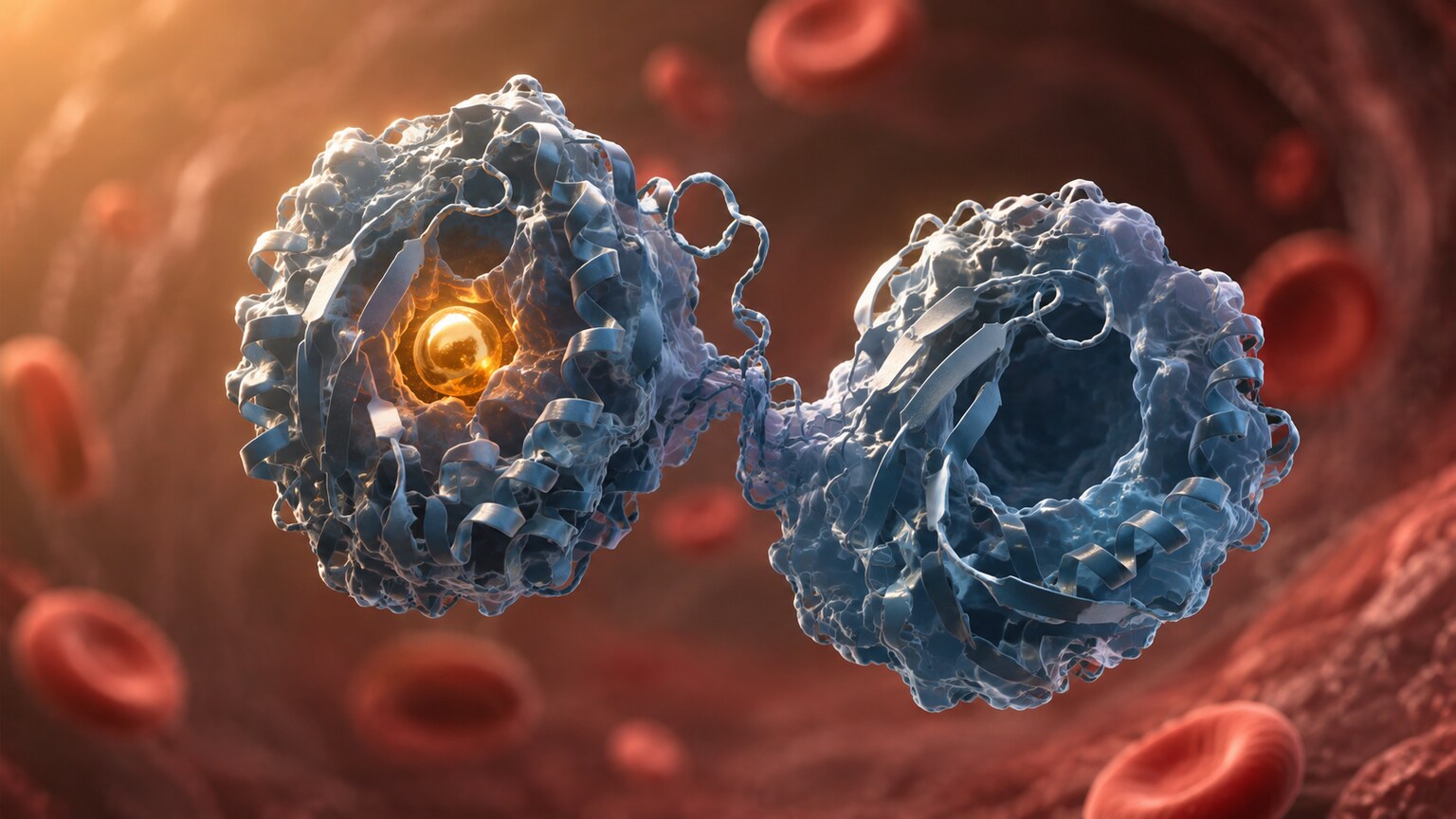
Figure 1. Each transferrin molecule has two binding sites capable of carrying an iron atom; TIBC reflects the total number of these sites across all transferrin molecules in a blood sample, not how many are actually occupied.
Already have a TIBC number from a recent iron panel, but not sure if it's actually good or bad for you? Just plug it in and see instantly.
🧮 Try the Free CalculatorTransferrin: The Molecule That Makes TIBC Possible
To understand TIBC, you first have to understand transferrin, the protein it's actually measuring. Transferrin is manufactured by the liver and released into the bloodstream, where its entire job is to bind free iron atoms and ferry them safely to wherever the body needs them — most often to the bone marrow, where iron gets built into new red blood cells. Free iron floating loose in the blood is chemically dangerous: it reacts readily with oxygen to generate damaging molecules called reactive oxygen species, and it's also a resource that many disease-causing bacteria actively try to steal for their own growth. Transferrin solves both problems at once, by keeping iron tightly bound and chaperoned everywhere it travels, essentially never letting it move through the bloodstream unescorted.
Each individual transferrin molecule has exactly two binding sites, meaning it can carry up to two iron atoms at a time, one at each end of its structure. In a healthy person, transferrin is typically only about 20% to 50% saturated with iron at any given moment — meaning most transferrin molecules in circulation are carrying either one iron atom or none at all, well short of their full two-atom capacity. This built-in slack matters enormously, because it means the body always keeps a meaningful reserve of unused iron-carrying capacity on hand, ready to absorb a fresh supply of iron the moment it becomes available from digestion or from the breakdown of old red blood cells. Transferrin also has a relatively short half-life in circulation, on the order of about eight to ten days, meaning the body is constantly recycling and replacing its transferrin supply — a turnover fast enough that TIBC can shift meaningfully within one to two weeks in response to a genuine change in iron status, but slow enough that it won't jump around from one day to the next the way same-day dietary intake can influence serum iron alone.
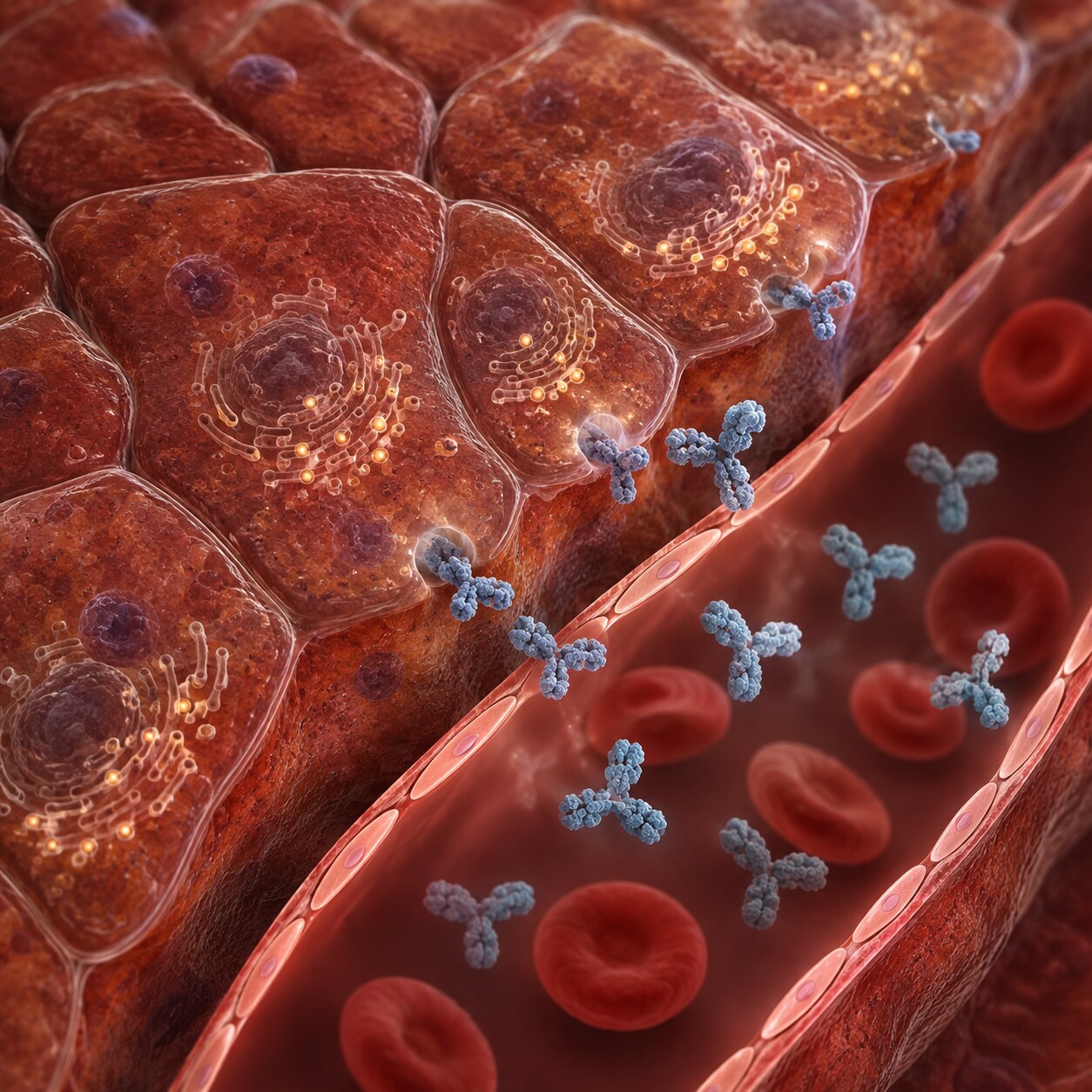
Figure 2. Transferrin is manufactured by the liver, which ramps production up or down in direct response to how much iron the body currently has available.
This is also where the test gets its name. In a lab, TIBC is measured by taking a blood sample and deliberately adding an excess of iron to it — far more than any transferrin present could possibly bind — which forces every single available binding site on every transferrin molecule to become fully saturated. The excess, unbound iron is then filtered out and discarded, and what remains is measured to determine exactly how much iron the sample's transferrin was capable of holding once fully loaded. That maximum-possible-binding number, expressed in micrograms of iron per deciliter of blood, is the total iron-binding capacity — a direct, physical measurement of transferrin quantity, expressed in the units of what it can carry rather than in units of protein mass directly.
Why TIBC Rises When Iron Stores Run Low
The relationship between TIBC and iron deficiency is one of the more elegant pieces of feedback biology in routine lab testing, and it's worth walking through carefully because it explains a pattern that confuses a lot of people at first glance: iron deficiency causing a lab value to go up rather than down.
The liver doesn't produce transferrin at a fixed rate — it adjusts production based on signals reflecting the body's current iron status, largely through a regulatory pathway involving a hormone called hepcidin, the body's master controller of iron balance. When the body's iron stores start running low, hepcidin levels drop, and this shift triggers the liver to ramp up transferrin production substantially, essentially manufacturing more transport capacity in anticipation of needing to capture and deliver whatever iron it can find. The logic mirrors something intuitive: if a shipping company suddenly has far less cargo to move, it doesn't necessarily buy more trucks — but the body's iron system does something closer to the opposite, expanding its fleet of transferrin carriers specifically because there's a shortage, as if trying to maximize its odds of catching every scarce iron atom passing through the bloodstream or absorbed from a meal.

Figure 3. On a printed iron panel, TIBC is typically listed alongside serum iron and transferrin saturation, and the three values are read together rather than in isolation.
This is precisely why TIBC and serum iron so often move in opposite directions on the same report: as iron deficiency develops, serum iron (the actual amount of iron circulating) falls, while TIBC (the amount of transport capacity) rises — the body has fewer iron atoms to carry, but more seats built to carry them. This combination — low serum iron paired with high TIBC — is one of the classic, textbook signatures of true iron-deficiency anemia, and it's specifically the contrast between the two numbers, not either one read alone, that gives a clinician real diagnostic confidence.
Transferrin Saturation: The Number That Ties Everything Together
Because TIBC on its own only tells you about capacity, not what's actually being carried, it's almost always interpreted alongside serum iron to calculate a third figure: transferrin saturation, sometimes abbreviated TSAT. This is simply serum iron divided by TIBC, multiplied by 100 to express it as a percentage — a direct answer to the question "of all the available seats, what percentage are currently filled?" A normal transferrin saturation typically falls somewhere between about 20% and 50%, meaning a healthy person's iron transport system runs with meaningful spare capacity built in, neither running nearly empty nor nearly maxed out under ordinary circumstances.
In iron deficiency, transferrin saturation drops well below that range, sometimes into the single digits in severe cases, because TIBC has risen (more empty seats) at the same time serum iron has fallen (fewer iron atoms to fill them) — a double effect on the same ratio, pushing the percentage down from both directions simultaneously. In iron overload conditions, the opposite dynamic tends to occur: TIBC often trends toward the lower end of normal or slightly below it, since the body isn't manufacturing extra transferrin when iron is already abundant, while serum iron itself may be elevated, together pushing transferrin saturation up toward or even past 45% to 50%. A transferrin saturation persistently above roughly 45% is one of the standard early screening signals clinicians watch for in hereditary hemochromatosis, a genetic condition that causes the body to absorb and store too much iron over time.
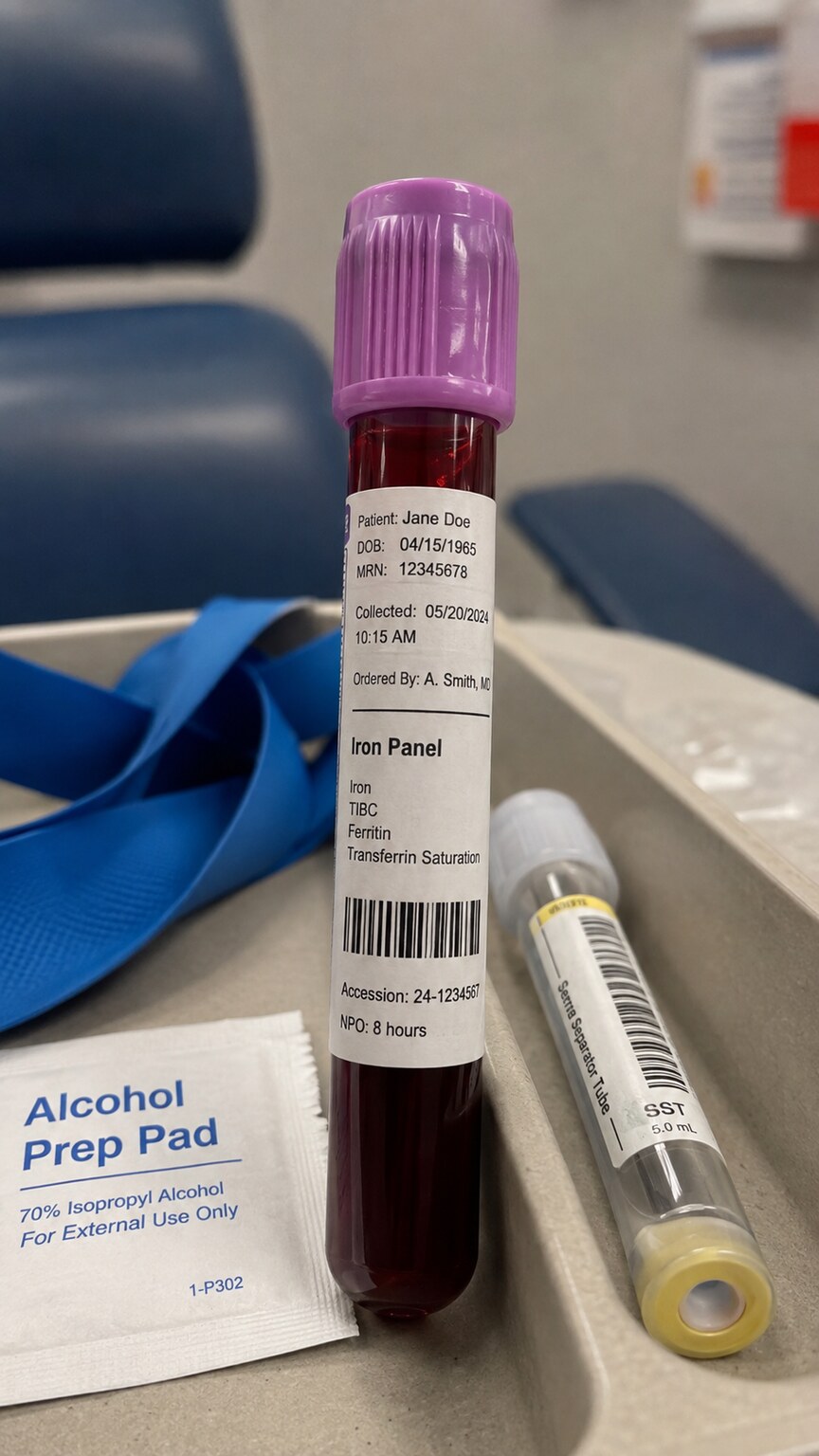
Figure 4. A single blood draw is typically enough to measure serum iron, TIBC, and ferritin together as part of a complete iron panel.
This is exactly why TIBC is rarely, if ever, ordered completely alone in modern practice — it's almost always drawn as part of a full iron panel that includes serum iron, transferrin saturation, and usually ferritin as well, precisely because each piece answers a different part of the same underlying question and none of them is reliable enough in isolation to stand on its own.
It's worth walking through a concrete example to make this concrete rather than abstract. Suppose a lab report shows serum iron of 40 mcg/dL (on the low side) and TIBC of 480 mcg/dL (above a typical upper limit of roughly 450). Dividing 40 by 480 and multiplying by 100 gives a transferrin saturation of about 8% — well below the normal 20% to 50% range, and low enough on its own to raise real concern for iron deficiency even before considering ferritin. Now compare that to a different report showing the same serum iron of 40 mcg/dL, but a TIBC of only 260 mcg/dL: the resulting transferrin saturation comes out to roughly 15%, still on the low side but nowhere near as alarming, and combined with a low TIBC rather than a high one, this second pattern points away from classic iron-deficiency anemia and toward something like anemia of chronic disease instead, where the body has plenty of transferrin capacity but inflammation is suppressing both iron mobilization and transferrin production together. The identical serum iron value, paired with two different TIBC results, tells two genuinely different clinical stories — which is the entire reason TIBC exists as a separate measurement rather than iron testing simply stopping at serum iron alone.
How TIBC, Ferritin, and Serum Iron Divide the Diagnostic Work
It helps to think of the three main iron-panel numbers as answering three distinct, complementary questions rather than three versions of the same question. Serum iron answers "how much iron is in the blood right this moment" — but this number swings significantly throughout the day, drops after a meal in some patterns and rises after others, and can shift meaningfully just from recent dietary intake or supplement timing, which makes it a fairly noisy, moment-in-time signal on its own. Ferritin answers a very different question: "how much iron is stored away in reserve," since ferritin is the body's primary iron-storage protein, mostly held inside the liver, spleen, and bone marrow rather than circulating loose in blood; a small, roughly proportional amount does leak into the bloodstream and gets measured on a standard test, making ferritin the most direct available window into total-body iron reserves under normal, non-inflamed conditions.
TIBC answers yet a third question: "how aggressively is the body trying to acquire more iron right now," since it reflects the liver's real-time manufacturing response to perceived iron shortage. Because TIBC responds directly to the body's own regulatory signaling rather than to momentary dietary fluctuations, it tends to be a steadier, more stable number day to day than serum iron alone, which is part of why the low-iron/high-TIBC/low-transferrin-saturation combination is considered such a reliable pattern for confirming true iron deficiency, especially when ferritin results are ambiguous.
Why the Body Evolved This Particular System
It's worth stepping back and asking why the body relies on this somewhat roundabout system — a dedicated carrier protein, adjustable in quantity, rather than simply letting iron circulate freely and adjusting absorption alone. The answer connects back to a genuinely ancient evolutionary pressure: nearly every disease-causing microorganism that has ever infected a vertebrate also needs iron to grow and reproduce, and many have evolved specific molecular machinery purpose-built to steal iron directly from their host's bloodstream. An animal that let iron circulate loose and unguarded would be handing invading bacteria an easily accessible fuel supply. Keeping essentially all circulating iron locked inside transferrin, accessible only through specific receptor proteins that the body itself controls, functions as a defense mechanism as much as a transport mechanism — a strategy immunologists sometimes call "nutritional immunity."
This dual purpose explains a pattern that would otherwise seem strange: during a serious infection, the body doesn't just fail to make more transferrin — it actively drives iron further into intracellular storage and tightens its grip on what little transferrin remains, specifically to starve invading pathogens of iron even at the cost of temporarily worsening the host's own red blood cell production. This is the deeper biological reason behind anemia of chronic disease and inflammation, discussed further below: it isn't simply an unfortunate side effect of illness, but appears to be, at least in part, a deliberate defensive tradeoff the body is making, sacrificing some red blood cell production in exchange for denying iron to whatever is making the person sick. Seen through that lens, TIBC isn't just a passive gauge of iron status — it's a readout of one of the body's active, ancient strategies for fighting infection, which is exactly why it behaves so differently in inflammatory illness than it does in straightforward nutritional iron deficiency.
Why Inflammation Complicates the Picture
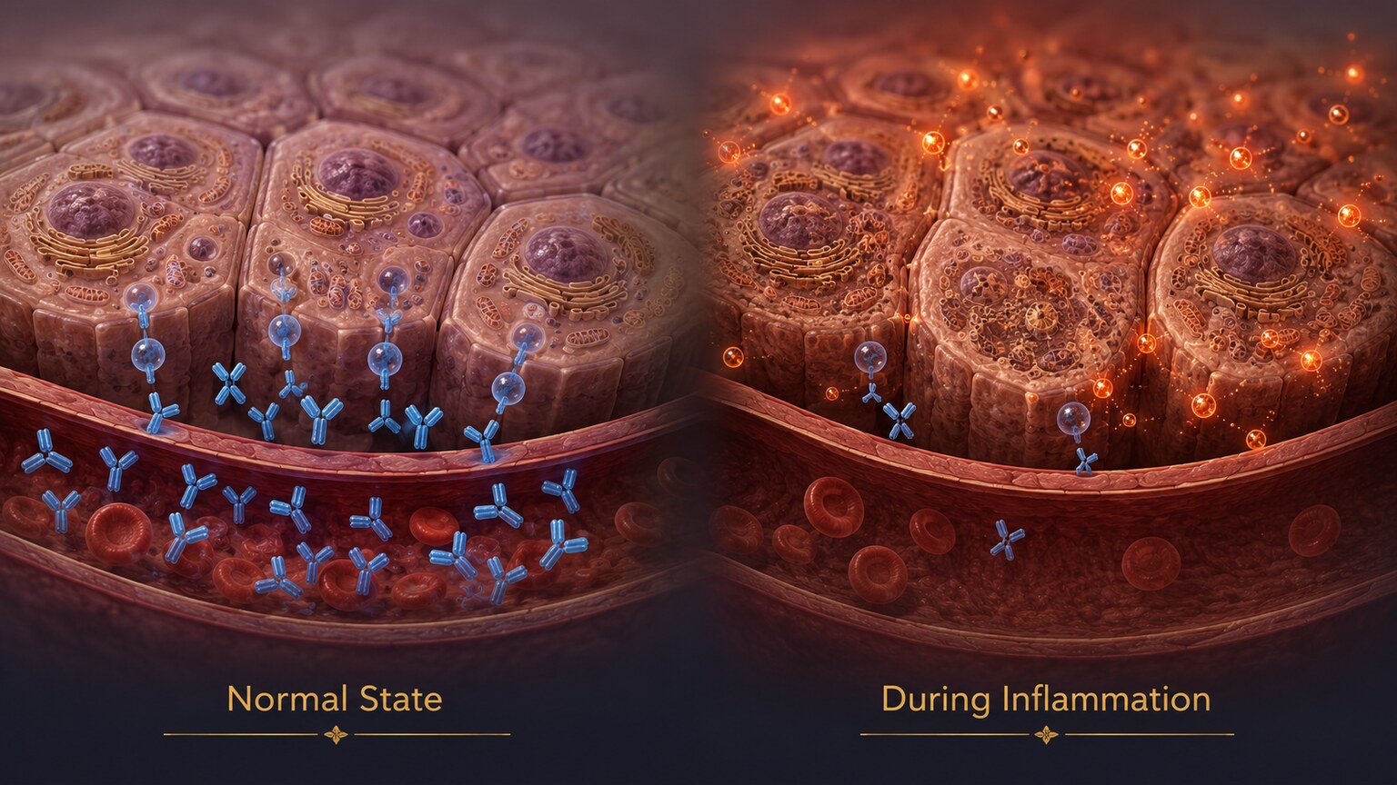
Figure 5. During active inflammation, the liver shifts priorities toward producing inflammatory proteins and away from transferrin, which can lower TIBC independently of a person's actual iron status.
Transferrin belongs to a broader category of proteins the liver produces called negative acute-phase reactants, meaning its production tends to decrease, not increase, during active inflammation, infection, or illness — the liver essentially reallocates its protein-manufacturing effort toward other proteins more directly useful for fighting infection or managing tissue damage in the short term, at transferrin's expense. This means TIBC can come back lower than expected in someone who is inflamed or acutely ill, even if that person's underlying iron stores are genuinely fine or even low — creating a scenario where inflammation can mask a real iron deficiency that would otherwise have shown up as an elevated TIBC.
This interaction is one of the most clinically important nuances of iron testing, and it's a major reason lab results from someone with a chronic inflammatory condition, a recent infection, or an autoimmune flare need to be interpreted with extra caution rather than taken at face value against standard reference ranges. In these situations, a clinician often leans more heavily on additional markers, such as inflammatory blood tests run alongside the iron panel, or considers a bone marrow evaluation in genuinely ambiguous or high-stakes cases, rather than relying on TIBC and ferritin numbers that inflammation may have distorted in opposite directions from what a person's true iron status would otherwise suggest.
What Counts as a Normal TIBC Range
TIBC results are reported in micrograms of iron per deciliter of blood (mcg/dL), and for most adults, a typical reference range runs somewhere between roughly 240 and 450 mcg/dL, though the exact cutoffs vary meaningfully from one laboratory to the next depending on the specific assay method and the reference population used to establish that lab's normal range. This is a detail worth taking seriously rather than glossing over: a TIBC of 410 mcg/dL might sit comfortably inside the normal range at one lab and land just outside the upper boundary at another, purely because of differences in how each lab calibrated its testing equipment and which population of healthy volunteers it used to define "normal" in the first place. This is exactly why every lab report prints its own reference range directly alongside the result, and why comparing a single number against a range remembered from a different lab, a different year, or a general internet search can be misleading.
It's also worth knowing that some labs report iron-binding results in a slightly different unit, micromoles per liter (mcmol/L), particularly outside the United States or in academic literature drawing on international studies — a conversion factor of roughly 0.179 translates mcg/dL to mcmol/L, and getting these units confused when comparing an old report to a new one, especially from a different country or health system, can make an otherwise perfectly normal result look dramatically abnormal at a glance.
Age and sex also shift what counts as typical. Children generally run somewhat higher TIBC values than adults, reflecting the elevated iron demands of active growth, while some studies have found modestly higher average TIBC in women than men outside of pregnancy, plausibly related to the additional iron losses menstruation represents across a lifetime. None of this means a single reference range is "wrong" — it means TIBC, like most lab values, is best interpreted in the context of the specific person being tested, not applied as a single universal cutoff to everyone regardless of age, sex, or physiological state.
What Causes TIBC to Be Abnormally Low
Beyond inflammation, a handful of other conditions can independently push TIBC toward the lower end of normal or below it, each through a distinct mechanism worth understanding on its own terms. Chronic liver disease is one of the more direct causes, since transferrin is manufactured in the liver — significant, longstanding liver damage from conditions like cirrhosis can impair the organ's overall protein-synthesis capacity broadly, transferrin included, independent of iron status entirely. Malnutrition, particularly a diet chronically low in overall protein rather than iron specifically, can similarly limit the raw material available for transferrin production.
Nephrotic syndrome, a kidney condition characterized by excessive protein loss through urine, causes transferrin to be lost from the body directly through the damaged kidney filtration system, lowering TIBC through a completely different route than reduced production — the protein is being made at a normal rate but escaping the body faster than usual. And as covered above, iron overload conditions themselves, including hereditary hemochromatosis and certain chronic transfusion-related iron accumulation, tend to produce TIBC values at or below the low end of normal, since an iron-replete body has no biological reason to manufacture extra transport capacity it doesn't currently need.
Certain medications can also lower TIBC independent of any of the mechanisms above. Corticosteroids, used to treat a wide range of inflammatory and autoimmune conditions, have been observed to reduce transferrin levels somewhat, likely tied to their broader effects on liver protein synthesis and their role in dampening the same inflammatory signaling pathways discussed throughout this article. L-asparaginase, a chemotherapy medication used primarily in the treatment of certain leukemias, has a more direct effect, since it specifically interferes with protein synthesis pathways in a way that can measurably lower TIBC during treatment. Neither of these medication effects is usually mistaken for iron overload once a clinician has a patient's full medication history in hand, but they're worth knowing about specifically because a TIBC drawn without that context could otherwise be misread.
What Causes TIBC to Be Abnormally High
On the other end, while iron deficiency is by far the most common driver of an elevated TIBC, it isn't the only one. Pregnancy, particularly in the second and third trimesters, is a well-documented cause of naturally rising TIBC independent of a woman's underlying iron stores, driven by estrogen's direct stimulating effect on the liver's transferrin production — which is part of why iron panel reference ranges used during pregnancy sometimes differ from standard adult ranges, and why an elevated TIBC in a pregnant patient isn't automatically assumed to represent a deficiency requiring the same workup it would outside of pregnancy. Estrogen-containing oral contraceptives and hormone replacement therapy have been shown to produce a similar, if generally milder, upward shift for the same underlying hormonal reason.
Because of these hormone-driven exceptions, a clinician interpreting an elevated TIBC always considers a patient's full context — reproductive status, medication list, and the pattern of the other iron panel values together — rather than assuming iron deficiency automatically explains every elevated result on its own.
A rare genetic condition called atransferrinemia is worth mentioning briefly, precisely because it illustrates just how central transferrin is to normal iron handling. People born with this condition produce little to no functional transferrin at all, and the consequences are severe: iron absorbed from the diet has no proper transport system to reach the bone marrow, leading to profound anemia despite iron actually accumulating, unused and mishandled, in other tissues like the liver and heart. While vanishingly rare, this extreme example underscores the broader point running through this entire article — TIBC isn't measuring a minor technical detail of blood chemistry, it's measuring the availability of an entire transport system the body depends on completely to move iron to where it's actually needed.
A Brief History of Why This Particular Test Exists
TIBC has been a routine part of clinical iron testing since the middle of the twentieth century, developed at a time when directly measuring transferrin protein levels was far more technically difficult and expensive than measuring what that protein could do — namely, how much iron it could bind once fully saturated. Rather than isolating and quantifying the transferrin molecule itself, early clinical chemists realized they could infer essentially the same information indirectly, by saturating a blood sample with excess iron and measuring how much of it got bound before the rest had to be removed. This indirect, functional approach turned out to be both cheaper and, in some ways, more clinically meaningful than a raw protein count would have been, since it captures transferrin's actual iron-carrying behavior rather than just its concentration.
Modern laboratories can now measure transferrin protein directly using immunoassay techniques, and where that's available, TIBC is sometimes calculated mathematically from the direct transferrin measurement instead of performed as the original iron-saturation method — the two approaches generally agree closely, since they're measuring the same underlying biological reality from two different technical angles. Despite newer, more direct methods existing, TIBC has remained the standard terminology and the standard way results are reported and interpreted across most of clinical medicine, largely because decades of published research, reference ranges, and clinical decision-making are all built around it, and switching terminology wholesale would create more confusion than it would resolve.
TIBC in Children, Pregnancy, and Chronic Disease
The way TIBC behaves — and the way it should be interpreted — shifts across different life stages and health conditions in ways worth understanding individually, since a single "normal adult range" doesn't apply equally well everywhere. In infants and young children, TIBC tends to run higher than adult reference ranges even at baseline, reflecting rapid growth and correspondingly elevated iron demand; pediatric iron-deficiency anemia, which remains common worldwide, particularly in toddlers transitioning off iron-fortified infant formula, typically shows the same elevated-TIBC, low-serum-iron pattern seen in adults, just measured against a different set of reference numbers appropriate for a child's age.
In pregnancy, beyond the estrogen-driven baseline rise already discussed, iron demand itself increases substantially to support the expanding blood volume and the developing fetus, meaning pregnant patients are simultaneously more prone to true iron deficiency and more likely to show an elevated TIBC from hormonal effects alone — a combination that makes the iron panel somewhat trickier to interpret during pregnancy than outside of it, and part of why routine iron supplementation is so commonly recommended during pregnancy regardless of borderline lab values, rather than waiting for a clearly abnormal result to intervene.
In chronic kidney disease, chronic inflammatory conditions like rheumatoid arthritis or inflammatory bowel disease, and cancer, the inflammation-driven suppression of TIBC discussed earlier becomes a persistent, ongoing feature rather than a temporary blip, which is part of why "anemia of chronic disease" is recognized as its own distinct diagnostic category, requiring a different interpretive lens than straightforward nutritional iron deficiency — the iron panel numbers in these patients can look confusingly similar to iron overload at a glance (low TIBC) while the person is actually functionally iron-restricted at the cellular level, unable to access or use the iron their body is actually storing.
How an Abnormal TIBC Result Shapes What Happens Next
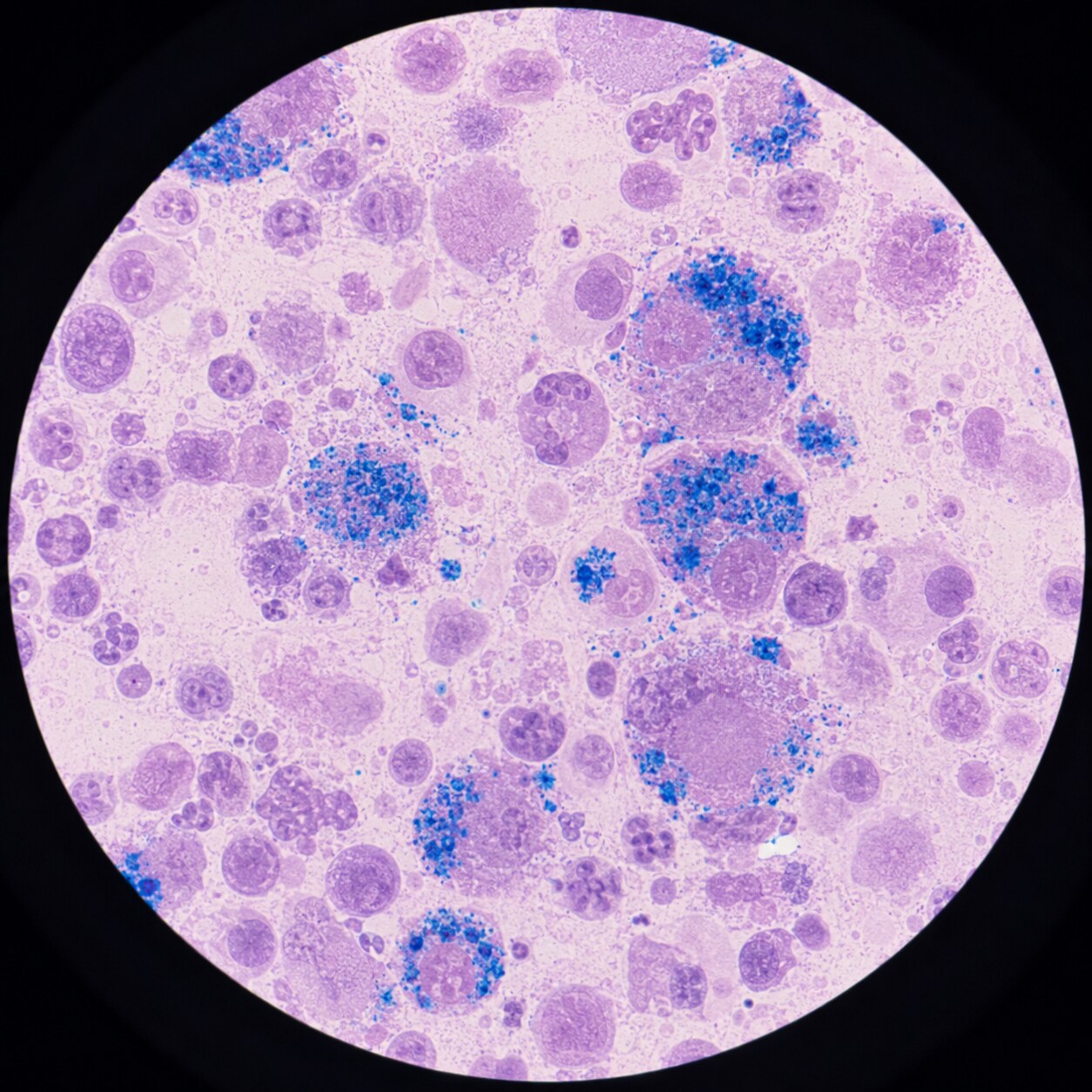
Figure 6. In genuinely ambiguous cases, particularly when inflammation may be distorting the iron panel, a bone marrow iron stain can serve as a more direct confirmation of the body's true iron reserves.
A clear pattern of low serum iron, high TIBC, and low transferrin saturation, in someone without significant active inflammation, is usually enough on its own for a clinician to confidently diagnose iron deficiency and move straight to identifying its cause — dietary insufficiency, heavy menstrual bleeding, gastrointestinal blood loss, or an absorption problem — rather than ordering further iron-specific testing. Treatment in that case typically means oral or, in some situations, intravenous iron replacement, followed by a repeat iron panel some weeks later to confirm the numbers are moving back toward normal.
When the picture is muddier — for instance, a low TIBC that doesn't fit cleanly with an otherwise iron-deficient clinical picture, or values that shift unpredictably alongside signs of active inflammation — a clinician may reach for additional tools rather than relying on the iron panel numbers alone. A soluble transferrin receptor test, which is less affected by inflammation than standard ferritin, is one such option gaining wider use specifically for these ambiguous cases. In the least clear or highest-stakes situations, a bone marrow aspirate stained specifically to reveal iron content remains the most direct, if far more invasive, way to confirm what the body's actual iron reserves look like, bypassing the indirect blood-protein signals entirely and observing the iron itself.
Frequently Asked Questions
Is a high TIBC always a sign of iron deficiency?
No. While iron deficiency is the most common cause of an elevated TIBC, pregnancy and estrogen-containing medications can also raise it through a hormonal pathway unrelated to how much iron the body actually has. A clinician typically looks at the full iron panel and clinical context together rather than treating an elevated TIBC alone as a diagnosis.
Why would my doctor order TIBC instead of just checking ferritin?
Ferritin and TIBC answer different questions — ferritin reflects stored iron reserves, while TIBC reflects how aggressively the body is trying to acquire more iron. They're most useful together, especially since inflammation can artificially lower TIBC and raise ferritin at the same time, making the combination more informative than either value alone in ambiguous cases.
Can TIBC be normal even with true iron deficiency?
It's uncommon but possible, particularly if inflammation is present at the same time, since inflammation tends to suppress transferrin production and can counteract the rise TIBC would otherwise show. This is one reason a single normal TIBC result doesn't always fully rule out iron deficiency in someone with a concurrent inflammatory condition.
Does fasting affect a TIBC test?
TIBC itself, since it measures transferrin's maximum capacity rather than current iron levels, is relatively stable and not strongly affected by recent food intake. However, because TIBC is almost always drawn alongside serum iron, which does fluctuate with meals and follows a daily rhythm, morning fasting blood draws are still generally recommended for the full iron panel to keep the paired serum iron result reliable.
How is TIBC different from UIBC?
UIBC, or unsaturated iron-binding capacity, measures only the currently empty seats on transferrin, while TIBC measures the total number of seats, filled and empty combined. The two are mathematically related — TIBC equals serum iron plus UIBC — and some labs report UIBC directly instead of, or alongside, TIBC, since either one combined with serum iron allows the same transferrin saturation percentage to be calculated.
Can diet or iron supplements change a TIBC result quickly?
Not meaningfully in the short term. Because TIBC reflects how much transferrin protein the liver has already manufactured, and that production process happens over days to weeks rather than hours, a single iron-rich meal or a few days of supplementation won't noticeably shift a TIBC result the way it can shift serum iron. Sustained dietary change or supplementation over several weeks is generally what's needed before a repeat TIBC would be expected to show meaningful improvement.
How TIBC Is Typically Presented on a Lab Report
Understanding what to actually look for on a printed or digital lab report can make the whole panel feel far less intimidating. TIBC is usually listed as its own line item, often positioned near serum iron and transferrin saturation within a section labeled "Iron Studies" or "Iron Panel," each with its own reference range printed alongside it and typically flagged with an "H" for high or "L" for low if the result falls outside that range. Some reports calculate and display transferrin saturation automatically as a fourth value, while others leave it to be calculated by hand or expect the ordering clinician to do the division themselves from the raw serum iron and TIBC numbers. If a report includes UIBC instead of, or in addition to, TIBC, the same underlying information is still there — as covered in the FAQ below, the two are directly related through simple subtraction, and either one paired with serum iron tells the same essential story about how full or empty the body's iron-transport capacity currently is.
Conclusion
TIBC is one of those lab values that only makes sense once you understand what it's actually built to measure — not iron itself, but the body's manufactured capacity to carry iron, a number that rises specifically because the body senses a shortage and responds by building more transport infrastructure. Read alone, it's a somewhat abstract figure; read alongside serum iron and ferritin, as part of a complete iron panel, it becomes one of the more reliable signals available for distinguishing true iron deficiency from other causes of anemia, and for catching a body's early attempt to compensate for a shortage before more familiar symptoms like fatigue or paleness ever show up. Understanding that TIBC measures capacity, not content, is the single fact that unlocks the rest of how it's used — and it's a distinction worth remembering the next time an iron panel comes back with a number that, at first glance, seems to be moving in exactly the wrong direction.
Still Not Sure What Your Results Mean?
Upload your labs and get a complete, visual, plain-language interpretation of every biomarker — delivered to your inbox in under 15 minutes.
Get My ReportThis article is for educational purposes only and does not constitute medical advice. Always consult your healthcare provider regarding your specific lab results.