
What's the Connection Between B12 and Anemia?
Low vitamin B12 and anemia are so consistently paired together in casual health conversation that it's easy to assume the connection is a simple, obvious one: not enough B12, so not enough red blood cells, essentially the same basic underlying story most people already associate with iron-deficiency anemia, just with a different vitamin swapped in as the missing ingredient. The actual underlying mechanism is meaningfully different from that assumption, though, and understanding it explains something that genuinely surprises a lot of people the first time they see their own results laid out in front of them: B12-deficiency anemia doesn't usually show up as a simple, straightforward low red blood cell count on its own — instead, it shows up as red blood cells that are the wrong size entirely, unusually and noticeably large, because B12's actual biological job in this process isn't simply to build more cells from scratch — it's to help each individual dividing cell finish accurately copying its own DNA before it can complete division. When that specific copying process stalls partway through, the bone marrow doesn't simply respond by making fewer cells overall; instead, it ends up producing fewer usable cells that are also noticeably larger and structurally defective compared to a normal, correctly matured red blood cell. This article walks through exactly why that happens at the cellular level, what the resulting pattern actually looks like once it shows up on a real lab report, and how doctors go about telling it apart from the handful of other conditions capable of producing a similarly enlarged red blood cell on the exact same test.

Figure 1. A B12-deficient red blood cell isn't simply reduced in number — it's abnormally enlarged, a direct consequence of DNA synthesis stalling mid-division inside the bone marrow.
Why B12 Deficiency Produces an Oversized Cell, Not Just Fewer Cells
Every red blood cell begins its life cycle inside the bone marrow as a much larger, immature precursor cell that has to divide several successive times, faithfully copying its full set of DNA at each individual division, before it finally shrinks down into the small, flexible, oxygen-carrying disc shape that eventually enters your circulating bloodstream and starts doing its actual job. Vitamin B12 is specifically required as a cofactor for an enzyme involved further along in producing thymidine, one of the four essential nucleotide building blocks that DNA itself is physically constructed from, meaning without it that step in the chain simply cannot proceed at its normal pace. Without enough B12, the cell can't manufacture enough thymidine to complete DNA synthesis on schedule.
Here's the specific, precise mismatch that defines this entire condition from start to finish: the cell's internal DNA-copying machinery stalls out partway through, but the rest of the cell — its surrounding cytoplasm, its overall physical growth and volume — keeps right on progressing largely unaffected in the meantime, since that particular ongoing process doesn't actually depend on adequate B12 availability in the same direct way the DNA-copying step does. The end result of this specific mismatch is a single cell that keeps growing steadily larger in overall physical size while its own nucleus lags noticeably behind, genuinely unable to finish completing its division on the normal expected schedule that a healthy cell would follow. This specific mismatched, out-of-sync pattern of growth is what pathologists and hematologists refer to as megaloblastic change, and it's the single defining, unmistakable feature that a trained observer can actually see and identify directly under a microscope — an abnormally large, immature-looking precursor cell in the bone marrow, and, once it's eventually released into general circulation while still abnormally oversized, an unmistakably large mature red blood cell circulating in the bloodstream itself, exactly as described.
There's a second, equally important consequence of this stalled division that explains why the anemia can become surprisingly severe relative to how the problem started. Many of these developing, DNA-compromised cells never make it out of the bone marrow at all — they're recognized internally as defective and destroyed before completing maturation, a process called ineffective erythropoiesis. This means the bone marrow, in a very real sense, can be working overtime, producing an even greater number of red blood cell precursors than normal in an attempt to compensate, and yet still releasing fewer usable mature cells into circulation than a healthy marrow would, since so many of that increased output never survive to be released. This combination — a hyperactive, crowded bone marrow paired with a paradoxically low final cell count in the bloodstream — is part of what makes megaloblastic anemia mechanically distinct from anemias caused by simple underproduction, and it also explains a related laboratory finding worth knowing about: because so many developing cells are being destroyed internally within the bone marrow itself, certain breakdown products — lactate dehydrogenase (LDH) and indirect bilirubin — often turn up elevated on bloodwork, a pattern that can look superficially similar to the cell destruction seen in certain other types of anemia, even though the underlying mechanism here is production failure rather than the bloodstream destruction of already-mature cells.
What This Looks Like on Your Actual Lab Report
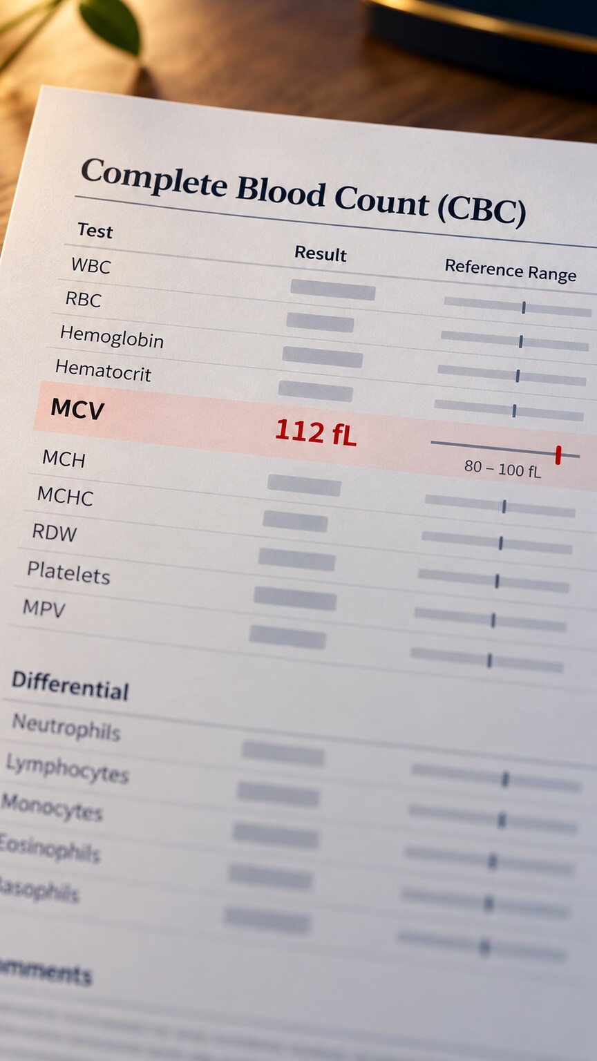
Figure 2. Mean corpuscular volume (MCV), the average size of a red blood cell, is typically the first and most direct clue on a routine CBC pointing toward B12-related megaloblastic anemia.
This mechanism translates directly into a specific, recognizable pattern on a standard complete blood count (CBC). Mean corpuscular volume, or MCV, measures the average size of your red blood cells, and B12 deficiency classically pushes this number above the normal range — a pattern called macrocytic anemia, meaning "large-celled" anemia. This is a genuinely useful distinguishing clue: iron-deficiency anemia, by contrast, typically produces the opposite pattern, a low MCV, since iron deficiency limits how much hemoglobin a cell can pack in, producing smaller, paler cells rather than larger ones. Seeing a low hemoglobin alongside a high MCV, rather than a low one, is often the first data point that points a doctor toward B12 or folate as the underlying cause rather than iron.
It's worth knowing the specific reference numbers involved, since they clarify exactly how a lab report communicates this pattern. Most laboratories define a normal MCV as roughly 80 to 100 femtoliters (fL), a unit describing the tiny volume of a single cell. A result climbing above 100 fL is what earns the "macrocytic" label, and B12 or folate deficiency severe enough to cause megaloblastic changes often pushes MCV considerably higher than that threshold, sometimes into the 110 to 130 fL range in more significant, longer-standing deficiency — a genuinely large cell by comparison, roughly a third larger in volume than a typical healthy red blood cell. This degree of enlargement is generally more extreme than the milder macrocytosis seen with some of the other, non-vitamin causes of an elevated MCV, which is itself a useful, if imperfect, clue a doctor can weigh when a very high MCV specifically raises suspicion for a vitamin deficiency over other possibilities.
Red cell distribution width, or RDW — a separate CBC value measuring how much size variation exists among a person's red blood cells rather than simply reporting the average — adds a further layer of useful detail to this picture. Early or mild B12 deficiency often shows an elevated RDW even before the average MCV itself has climbed clearly into the abnormal range, since the earliest affected cells becoming oversized initially just widen the overall size distribution rather than immediately dragging the whole average upward. This makes a rising RDW, tracked over serial testing, sometimes the very first hint of a developing problem — a subtle early warning sign that can appear on a routine CBC well before the anemia or the macrocytosis itself becomes obvious enough to draw attention on its own.
Curious what your own CBC or B12 results actually mean together? Upload your results and get a complete, plain-language breakdown in under 15 minutes.
Analyze My ResultsWhy Folate Almost Always Comes Up in the Same Conversation
Folate (vitamin B9) shares this exact mechanism with B12, which is precisely why the two are so often tested together and why a folate deficiency produces a laboratory picture that's essentially indistinguishable from B12 deficiency on a basic CBC. Folate is required at a different, earlier step of the very same thymidine-production pathway that B12 supports — the two vitamins work in sequence on the same overall process, meaning a shortage of either one stalls DNA synthesis in the same characteristic way and produces the same megaloblastic, macrocytic pattern.
This is exactly why a standalone elevated MCV can't tell a doctor which of the two vitamins is actually responsible without further testing, and why both are typically checked together whenever one is suspected. There's also a clinically important, occasionally dangerous interaction between the two worth understanding: high-dose folate supplementation can correct the anemia caused by an underlying B12 deficiency, since folate can partially substitute for B12 in this specific pathway — but it does nothing to address B12's separate, unrelated role in maintaining the protective coating around nerve cells. This means someone could see their anemia improve on folate alone while an underlying B12 deficiency continues silently causing nerve damage in the background, undetected until it becomes significant — a genuine reason doctors are cautious about high-dose folate supplementation without first confirming B12 status.
This concern isn't purely theoretical — it's directly reflected in food fortification policy in several countries, including the United States, where synthetic folic acid is added to enriched grain products specifically to reduce the incidence of neural tube birth defects. This same policy has a documented side effect worth understanding: it can effectively mask the anemia component of a B12 deficiency in the general population, since ambient folic acid intake from fortified foods is often enough to keep red blood cells closer to normal size even in someone with a genuine, ongoing B12 shortfall. This masking effect means the anemia and macrocytosis this article has described can be blunted or absent in some people with real B12 deficiency, while the separate nerve-related consequences of that same deficiency continue progressing unmasked and undetected in the background, since folate does nothing to protect the myelin-maintenance role that belongs to B12 alone. This is one of the more counterintuitive, less widely appreciated aspects of the B12-anemia relationship: in a population with widespread folic acid fortification, a normal-looking blood count provides measurably less reassurance about someone's true underlying B12 status than it would have a century ago, before fortification existed.
Pernicious Anemia: The Specific Autoimmune Cause Behind Many Cases

Figure 3. Pernicious anemia results from an autoimmune attack on the stomach cells that produce intrinsic factor, a protein required for B12 to be absorbed at all, regardless of how much is consumed in the diet.
The specific term "pernicious anemia" refers to one particular cause of B12-deficiency anemia, not the general condition itself, and it's worth knowing the distinction. B12 can't simply diffuse across the intestinal wall on its own — it needs to bind to a protein called intrinsic factor, produced by specific cells lining the stomach, in order to be recognized and absorbed further down in the small intestine. Pernicious anemia occurs when the immune system mistakenly produces antibodies against these stomach cells (or against intrinsic factor itself), destroying the body's ability to absorb B12 no matter how much is present in the diet.
This autoimmune mechanism explains why pernicious anemia can develop even in someone eating a diet rich in B12-containing foods like meat, eggs, and dairy — the problem isn't intake, it's absorption, and no amount of dietary correction fixes a broken absorption pathway. Pernicious anemia also tends to develop gradually over years, since the body's B12 stores in the liver are substantial and can take three to five years to fully deplete once absorption stops, which is part of why it's more commonly diagnosed in older adults and why symptoms can feel like they "came out of nowhere" despite years of underlying, silent depletion. Diagnosis of this specific condition typically involves testing directly for intrinsic factor antibodies, and once confirmed, treatment requires B12 delivered by a route that bypasses the broken absorption step entirely — injections, high-dose oral supplementation absorbed passively without intrinsic factor, or a nasal spray — rather than standard dietary or low-dose oral approaches.
It's worth understanding why intrinsic factor is genuinely irreplaceable in this process rather than simply one of several helpful absorption aids. Dietary B12 arrives in food bound to protein, and it first has to be released from that protein by stomach acid and a digestive enzyme before it can bind to intrinsic factor at all — meaning conditions that reduce stomach acid, including long-term use of certain acid-suppressing medications, can also modestly impair this earlier release step even when intrinsic factor production itself remains completely normal, producing a milder form of absorption-related deficiency through a different point of failure in the same overall pathway. Once properly bound, the B12-intrinsic factor complex travels the entire length of the small intestine essentially untouched, since it needs to reach highly specific receptors located only in the final segment, called the terminal ileum, where actual absorption into the bloodstream finally takes place. This explains a related but distinct cause of B12 deficiency worth mentioning alongside pernicious anemia: surgical removal of the terminal ileum, whether for Crohn's disease, cancer, or another condition, removes the only site in the entire digestive tract where this absorption step can occur, producing a B12 deficiency through anatomical loss of the necessary tissue rather than an autoimmune attack on it.
The autoimmune process behind pernicious anemia also frequently doesn't occur in isolation, a detail worth being aware of since it often prompts a broader evaluation once diagnosed. Pernicious anemia is strongly associated with other autoimmune conditions, particularly autoimmune thyroid disease, and to a lesser extent type 1 diabetes and vitiligo, reflecting a broader tendency for autoimmune conditions to cluster together in the same individual, likely related to shared underlying genetic and immune-regulatory factors that aren't yet fully understood. Because of this recognized clustering, a new diagnosis of pernicious anemia often prompts screening for thyroid dysfunction specifically, even in the absence of any thyroid-related symptoms, since an existing autoimmune process affecting one gland meaningfully raises the statistical likelihood of another developing at some point.
Beyond the Red Blood Cells: Why Other Cell Lines Are Affected Too
Because the underlying defect is in DNA synthesis generally, not something specific to red blood cells alone, B12 deficiency can also affect other rapidly dividing cell populations in the bone marrow, producing a broader pattern than anemia by itself. White blood cell counts can drop, and specifically, a distinctive finding called hypersegmented neutrophils — white blood cells whose nucleus has an unusually high number of lobes — is one of the earliest and most specific microscopic clues pointing toward megaloblastic anemia, sometimes appearing on a blood smear even before the red blood cells themselves become visibly enlarged. Platelet counts can drop as well, since platelet precursors divide rapidly too and are subject to the same DNA-synthesis bottleneck. This broader pattern — anemia, low white blood cells, and low platelets together — is sometimes called pancytopenia, and its presence alongside macrocytic red cells is a strong combined signal pointing toward B12 or folate deficiency specifically, rather than a more isolated cause of anemia.
Hypersegmented neutrophils deserve a slightly closer look, since the finding is specific enough to be genuinely useful on its own. A normal neutrophil's nucleus typically has three to five distinct lobes; a hypersegmented one shows six or more, a change that reflects the same underlying DNA-synthesis defect described throughout this article, just expressed in a different cell line than red blood cells. Because neutrophil precursors have a considerably shorter developmental timeline in the bone marrow than red blood cell precursors do, this particular abnormality can actually appear on a blood smear before the red blood cells themselves have had enough time to show clear megaloblastic changes, making it, in some cases, the single earliest visible clue a skilled technician can spot under the microscope, sometimes days to weeks before the anemia itself becomes laboratory-apparent.
The bone marrow findings in more severe or advanced cases can be dramatic enough to occasionally cause diagnostic confusion in their own right, which is worth understanding to avoid unnecessary alarm. A bone marrow biopsy performed in someone with significant, untreated megaloblastic anemia can show a strikingly hypercellular marrow — packed with far more developing cells than a normal marrow contains, reflecting the compensatory overproduction described earlier — alongside abnormal-looking, oversized precursor cells. This specific combination has, in some documented cases, been initially mistaken for a bone marrow malignancy like leukemia by clinicians unfamiliar with the pattern, before B12 and folate levels were checked and the true, entirely benign and reversible cause identified. This is precisely why current clinical guidance emphasizes checking B12 and folate levels early in the workup of any unexplained macrocytic anemia or unusual bone marrow finding, specifically to rule out this readily treatable explanation before pursuing more invasive or alarming diagnostic paths.
How Doctors Confirm the Diagnosis Beyond a Basic CBC

Figure 4. A hypersegmented neutrophil, with an unusually high number of nuclear lobes, is one of the earliest microscopic clues pointing toward megaloblastic anemia, sometimes visible before red cells themselves enlarge.
Because both B12 and folate deficiency, and even some non-vitamin causes, can produce a similar-looking macrocytic pattern, a doctor confirming the actual cause typically orders serum B12 and folate levels directly, alongside a peripheral blood smear to check for the specific features described above (oversized cells, hypersegmented neutrophils). When B12 comes back in a borderline or ambiguous range, a more sensitive follow-up test measuring methylmalonic acid (MMA) is often used — MMA reliably accumulates when B12 is genuinely deficient at the cellular level, since B12 is required to clear it, making an elevated MMA a more definitive confirmation than a borderline serum B12 number alone. Homocysteine, yet another compound that the body clears with direct help from both B12 and folate together, is likewise sometimes checked for this same general reason, though it's considerably less specific to B12 deficiency alone, since a folate deficiency raises this same homocysteine value too.
If pernicious anemia specifically is suspected, intrinsic factor antibody testing follows, and in some cases, particularly where the diagnosis remains unclear, an endoscopy examining the stomach lining directly can confirm the characteristic tissue changes associated with the autoimmune destruction of acid- and intrinsic-factor-producing cells.
It's worth understanding a limitation of the standard serum B12 test itself, since it explains why doctors sometimes pursue further testing even when the initial B12 number looks reassuringly normal. The standard blood test measures total circulating B12, but a meaningful fraction of that total is bound to a carrier protein in a form that isn't actually biologically usable by cells — meaning someone can have a technically normal total B12 result while still having a functional deficiency at the cellular level, since the usable fraction specifically is what's running low. This is part of why methylmalonic acid testing, mentioned earlier, carries particular value in ambiguous cases: because MMA reflects what's actually happening inside cells rather than the total amount circulating in blood, it can catch a genuine functional deficiency that a borderline-normal total B12 result would otherwise miss entirely, which is exactly the scenario where relying on a single test in isolation risks a missed or delayed diagnosis.
Age adds a further layer of complexity worth knowing about, since standard B12 reference ranges aren't perfectly calibrated for every population. Older adults have a higher background rate of reduced stomach acid production and mild, food-bound B12 malabsorption unrelated to pernicious anemia specifically, simply as a consequence of normal aging changes to the stomach lining over time. This means a borderline-low B12 result in an older adult carries a meaningfully higher pre-test likelihood of representing a genuine, clinically relevant deficiency than the identical result would in a younger person, which is part of why some clinicians apply a somewhat lower threshold for further workup or empiric treatment in this specific age group rather than treating the same numeric cutoff identically across every age.
Why Symptoms Can Appear Before the Anemia Is Severe

Figure 5. Because B12 supports both red blood cell production and nerve health through separate mechanisms, fatigue and pallor from anemia can appear alongside early tingling or numbness from nerve involvement.
Because B12 plays two largely separate biological roles — supporting DNA synthesis in dividing cells, and maintaining the protective myelin sheath around nerve fibers — its deficiency can produce two distinct categories of symptoms that don't always progress in lockstep with each other. The anemia-related symptoms are what you'd expect from any reduced oxygen-carrying capacity: fatigue, noticeable paleness, shortness of breath on exertion, and a rapid heartbeat as the body compensates. But nerve-related symptoms — tingling or numbness in the hands and feet, difficulty with balance, and in more advanced cases, memory or cognitive changes — stem from the separate myelin-related mechanism and can sometimes appear even before the anemia itself becomes severe enough to cause noticeable fatigue, particularly in cases where the deficiency has been developing slowly over years.
This decoupling matters clinically because it means a normal or only mildly reduced hemoglobin doesn't rule out a clinically significant B12 problem if nerve-related symptoms are already present — the two systems can be affected at different rates in different people, for reasons that aren't fully understood. It's part of why a doctor evaluating unexplained tingling or balance problems will often check B12 levels even when a basic CBC looks reassuringly close to normal, since waiting for the anemia to become obvious risks allowing nerve damage to progress further in the meantime.
How Doctors Track Whether Treatment Is Actually Working

Figure 6. A rising reticulocyte count within the first week of B12 treatment — the "reticulocyte response" — is the earliest reliable confirmation that the bone marrow has resumed normal red blood cell production.
Once treatment starts, doctors don't simply wait for hemoglobin to return to normal before confirming it's working, since that recovery can take weeks to fully complete. Instead, one of the earliest and most reliable indicators is a reticulocyte count — a measurement of newly released, slightly immature red blood cells — checked five to seven days after treatment begins. A properly functioning bone marrow, once supplied with adequate B12, responds by ramping up production quickly, producing a measurable spike in reticulocytes well before the overall hemoglobin has caught up, since it takes time for that surge of new cells to meaningfully raise the total count. This spike, called the reticulocyte response, is a genuinely reassuring early signal that the underlying mechanism has been correctly identified and corrected, and its absence within the expected window prompts a doctor to reconsider the diagnosis or look for a coexisting problem — such as concurrent iron deficiency, which can blunt the expected response even when B12 itself has been adequately replaced.
Serum B12 and MCV are typically rechecked over the following one to three months to confirm the trend continues in the right direction, though it's worth knowing that MCV can occasionally rise slightly further in the very first days of treatment before beginning to fall, as the bone marrow's rapid new production temporarily includes a wave of larger reticulocytes — a pattern that can look alarming out of context but is a normal, expected part of recovery rather than a sign that treatment isn't working.
A Worked Example: Two Similar-Looking Results, Two Different Explanations
Consider, first, a 68-year-old with fatigue and a CBC showing hemoglobin at 9.8 g/dL and an MCV of 112 fL (well above the typical upper limit near 100). Her serum B12 comes back low, and her doctor orders intrinsic factor antibodies, which return positive, confirming pernicious anemia. Treatment begins with B12 injections, bypassing her broken absorption pathway entirely, and her hemoglobin and MCV both gradually normalize over the following months as her bone marrow, now supplied with adequate B12, resumes producing correctly sized cells.
Now, by contrast, consider a 24-year-old with a similar CBC pattern — low hemoglobin, elevated MCV — who follows a strict vegan diet with no B12 supplementation. Her serum B12 also comes back low, but her intrinsic factor antibodies are negative, and her absorption is otherwise intact; the deficiency traces simply to inadequate dietary intake, since B12 occurs naturally almost exclusively in animal products. Her treatment is considerably simpler: oral B12 supplementation or fortified foods, without the need for injections, since her absorption pathway itself was never impaired. Two patients, a nearly identical lab picture, and two meaningfully different causes requiring two different treatment approaches — exactly why confirming the specific underlying cause matters as much as recognizing the pattern itself.
A third scenario illustrates why the interpretation gets more layered still. A 45-year-old with a history of gastric bypass surgery several years earlier presents with fatigue and a similar macrocytic pattern on her CBC. Her surgery altered both her stomach's acid- and intrinsic-factor-producing capacity and bypassed a portion of her small intestine, meaning her B12 deficiency traces to a surgically altered anatomy rather than either autoimmune destruction or simple dietary insufficiency — a third distinct mechanical category alongside the two already described. Her intrinsic factor antibodies come back negative, ruling out pernicious anemia specifically, but because her surgery has permanently altered how much intrinsic factor her remaining stomach tissue can produce, dietary correction alone won't reliably fix the problem long-term. Her treatment settles on regular B12 injections, similar to the approach used for pernicious anemia, not because she has that specific autoimmune condition, but because her particular anatomical change produces a comparably persistent absorption barrier that oral supplementation alone is unlikely to fully overcome — a reminder that the same practical treatment can apply to genuinely different underlying causes once the shared end result (an absorption pathway that can't be relied upon) is the same.
Frequently Asked Questions
Can B12-deficiency anemia be reversed?
Yes, in most cases. Once adequate B12 is restored, whether by injection, oral supplementation, or dietary correction depending on the cause, the bone marrow generally resumes producing normally sized red blood cells within weeks to a few months.
Is a high MCV always caused by B12 or folate deficiency?
No. Other causes of macrocytic anemia include certain medications, excessive alcohol use, liver disease, and some bone marrow disorders, which is why B12 and folate levels are checked directly rather than assumed from an elevated MCV alone.
Why do I need injections instead of just taking a B12 pill?
Injections are typically used when the underlying cause involves an absorption problem, like pernicious anemia, since injected B12 bypasses the digestive tract and broken intrinsic factor pathway entirely, unlike a standard oral supplement.
Can vegetarians and vegans avoid B12-deficiency anemia?
Yes, with appropriate supplementation or fortified foods. Since B12 occurs naturally almost exclusively in animal products, anyone on a strict plant-based diet needs a reliable alternative source to prevent deficiency from developing over time.
Does B12-deficiency anemia cause permanent damage if untreated?
The anemia itself typically resolves fully with treatment, but B12 deficiency can also cause nerve damage that, if left untreated long enough, may not fully reverse — a separate reason early diagnosis and treatment matter beyond correcting the blood count alone.
Can a normal B12 blood test still miss a real deficiency?
Yes, in some cases. Standard testing measures total circulating B12, which includes a biologically unusable fraction, so methylmalonic acid testing is sometimes used to catch a genuine functional deficiency that a borderline-normal total B12 result would otherwise miss.
How quickly does treatment start improving symptoms?
A rising reticulocyte count, an early sign the bone marrow is responding, typically appears within five to seven days of starting treatment, though full normalization of hemoglobin and resolution of fatigue generally takes several weeks to a few months.
Conclusion
The connection between B12 and anemia isn't a simple story of "not enough vitamin, not enough blood" — it's a specific breakdown in DNA synthesis that leaves red blood cells oversized and dysfunctional rather than merely scarce, a pattern visible on a routine CBC as an elevated MCV long before most other clues appear. Whether the underlying cause is dietary insufficiency, a surgically altered digestive anatomy, or the autoimmune destruction seen in pernicious anemia, confirming exactly which one applies is what determines whether the right fix is a dietary change, an oral supplement, or a lifelong injection routine — three genuinely different paths that all happen to produce the same abnormal number on the same test.
The broader takeaway worth carrying forward is that an elevated MCV, on its own, is really an invitation to ask a more specific question rather than an answer in itself. It narrows the field considerably — pointing away from iron deficiency and toward a shortage of B12, folate, or one of the less common alternative explanations — but the actual mechanism, the specific cause, and the correct fix only become clear once the follow-up testing described throughout this article fills in the rest of the picture. Treated that way, a single abnormal cell size on a routine blood count becomes a genuinely useful diagnostic thread to pull, rather than either a source of alarm or a number safely ignored.
Still Not Sure What Your Results Mean?
Upload your labs and get a complete, visual, plain-language interpretation of every biomarker — delivered to your inbox in under 15 minutes.
Get My ReportThis article is for educational purposes only and does not constitute medical advice. Always consult your healthcare provider regarding your specific lab results.