
What Does a High CA 19-9 Level Mean?
Here's a fact that surprises almost everyone the first time they hear it: the single highest CA 19-9 level ever documented for a genuinely benign condition — a simple bile duct stone, nothing malignant at all — was 187,000 U/mL. No case of pancreatic cancer in the medical literature has ever been recorded above 100,000 U/mL. In other words, at the very top of the range, an entirely benign result has outscored every malignant one on record. That single fact tells you something important: "high" isn't one thing. A CA 19-9 of 45 U/mL, 450 U/mL, and 45,000 U/mL are three genuinely different clinical situations, not three flavors of the same bad news. This article walks through the actual magnitude scale — the real cutoffs doctors use, what each range statistically suggests, and why the number climbing higher matters more in some ranges than others.
It helps to think of CA 19-9 less like a switch that flips from "normal" to "abnormal," and more like a dial with several distinct zones on it, each backed by its own body of published research and its own clinical response. Understanding which zone a specific result falls into — and what oncologists have actually found tends to be true in that zone — turns a single unfamiliar number into something you can reason about with real context, rather than a source of open-ended anxiety, before your next conversation with the person who ordered the test in the first place.
The Reference Range Is Just the Starting Line
Most labs flag anything above 37 U/mL as "elevated," but that single line does far less work than people assume. It marks the edge of the statistical range built from healthy people — not a boundary between "fine" and "cancer." Crossing it by even a small amount puts you in the same flagged category as someone whose result is a thousand times higher, even though those two situations mean almost nothing alike. Everything that follows in this article is really about what happens as you move further past that initial line, and how doctors think differently about each stretch of the climb.
It's worth noting upfront that 37 U/mL itself isn't a universal constant either — the exact cutoff can shift slightly depending on which assay a specific lab uses to run the test, and different cancer types have their own separately validated cutoffs that don't always match the general 37 U/mL figure most people encounter first. That variability is itself a clue that this marker was never designed to be read as a single, context-free pass/fail line, in any disease it's used for, which is exactly the idea the rest of this article unpacks range by range.

37 to 100 U/mL: Mildly Elevated Territory
This is by far the most common range for a flagged CA 19-9 to land in, and it's also the range where benign explanations dominate the statistics most heavily. Research has found that a median CA 19-9 under 100 U/mL correlates with resectable, earlier-stage disease in the minority of cases where cancer is actually present at all — and given how common benign causes are at this level, a result in this band is, on its own, one of the least alarming versions of an "abnormal" CA 19-9 a person can receive. It's also the range most likely to reflect one of the benign mechanisms covered in our companion article on what causes CA 19-9 to rise in benign conditions — mild pancreatitis, early cholestasis, poorly controlled diabetes, or simple day-to-day biological variation.
This is also the range where a repeat test earns the most value for the least anxiety. Because so many benign, temporary factors can nudge a result from, say, 34 to 55 U/mL — a recent illness, mild dehydration, a brief flare of an unrelated inflammatory condition — a single result modestly above 37 is often treated by doctors as a prompt to simply check again in a few weeks rather than an immediate trigger for extensive imaging. If the repeat draw settles back toward normal, that alone is meaningful reassurance; if it holds steady or climbs, that pattern is what actually starts to justify a closer look, not the single original number in isolation.
100 to 300 U/mL: Where Suspicion Starts to Build
Crossing 100 U/mL is a meaningful psychological and statistical threshold. Research has found that levels above 100 U/mL are associated with advanced or metastatic pancreatic cancer in roughly 60% to 85% of cases where a malignancy is ultimately confirmed — a sharp jump from the picture below 100. This is also the range where cancer-specific cutoffs for other digestive cancers start to matter: a cutoff of 46 U/mL has been established as optimal for ampullary cancer specifically, and a cutoff of 103 U/L for cholangiocarcinoma, above which metastasis and reduced survival become significantly more likely. Even within this band, though, benign causes haven't been ruled out — this is simply the range where a doctor starts taking the possibility of malignancy more seriously and typically orders imaging, if it hasn't been done already.
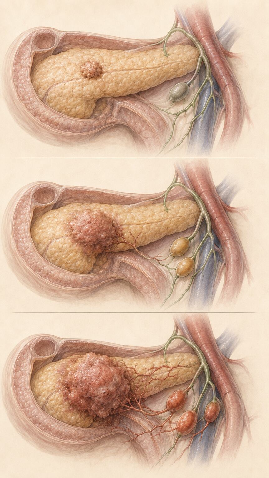
One specific finding worth knowing: among patients with confirmed pancreatic tumors, a CA 19-9 above 37 U/mL was associated with lymph node involvement in 75.9% of cT1 (smallest) tumors, 81.3% of cT2 tumors, and 85.7% of cT3 tumors — meaning the marker's association with spread beyond the primary tumor gets stronger, not weaker, as the tumor stage advances, even within this relatively modest numeric range. That climbing percentage, tied to tumor stage rather than to the CA 19-9 number alone, is a good early illustration of a theme that runs through the rest of this article: magnitude and biology are related, but the relationship isn't a simple straight line.
It's worth being precise about what "lymph node involvement" actually changes for a patient, since the phrase can sound more frightening in the abstract than the clinical reality always warrants. Lymph nodes are small, bean-shaped structures scattered throughout the body that filter fluid and house immune cells, and cancer cells sometimes travel to nearby ones before spreading further. Finding cancer in nearby lymph nodes generally shifts a tumor into a more advanced stage and changes the recommended treatment approach, often adding chemotherapy before or after surgery, but it does not automatically mean the disease is untreatable or that surgery is off the table entirely — it simply means the surgical and oncology team need a fuller picture of exactly how far the disease has traveled before finalizing a plan.
300 to 1,000 U/mL: Resectability Becomes the Central Question
Once a confirmed or strongly suspected pancreatic cancer sits in this range, the conversation among oncologists and surgeons shifts specifically toward whether the tumor can realistically be removed. A cutoff of 336 U/mL has been shown in meta-analysis to predict metastatic disease with 90% sensitivity and 80% specificity — strong enough performance that some surgical teams weigh it directly in staging discussions. Other studies have proposed a preoperative cutoff of 150 U/mL carrying an 88% positive predictive value for unresectability, and a cutoff around 178 U/mL described as strongly suggestive of unresectable disease. An international consensus has separately settled on 500 U/mL as one of the biological criteria used to classify a pancreatic tumor as "borderline resectable" — a formal staging category between clearly operable and clearly inoperable, used specifically because a marker in this zone often lines up with tumors that are anatomically borderline as well.
The existence of several different proposed cutoffs in this same general range — 150, 178, 336, 500 — isn't a sign that researchers can't agree on a simple fact. It reflects the reality that "resectable" isn't a single yes-or-no biological state either; it's a judgment call that weighs tumor size, its relationship to nearby blood vessels, whether nearby lymph nodes are involved, and a patient's overall fitness for major surgery, alongside the CA 19-9 number. Different research groups, studying somewhat different patient populations with somewhat different surgical thresholds, have understandably landed on somewhat different optimal cutoffs for their own specific decision points — which is exactly why your own surgical team's judgment, informed by their own imaging and staging workup, carries more weight for your specific case than any single published number from this article ever could.
Curious what your own lab results are really telling you? Upload them and get a complete, plain-language breakdown in under 15 minutes.
Analyze My ResultsDifferent Cancers, Different Cutoffs
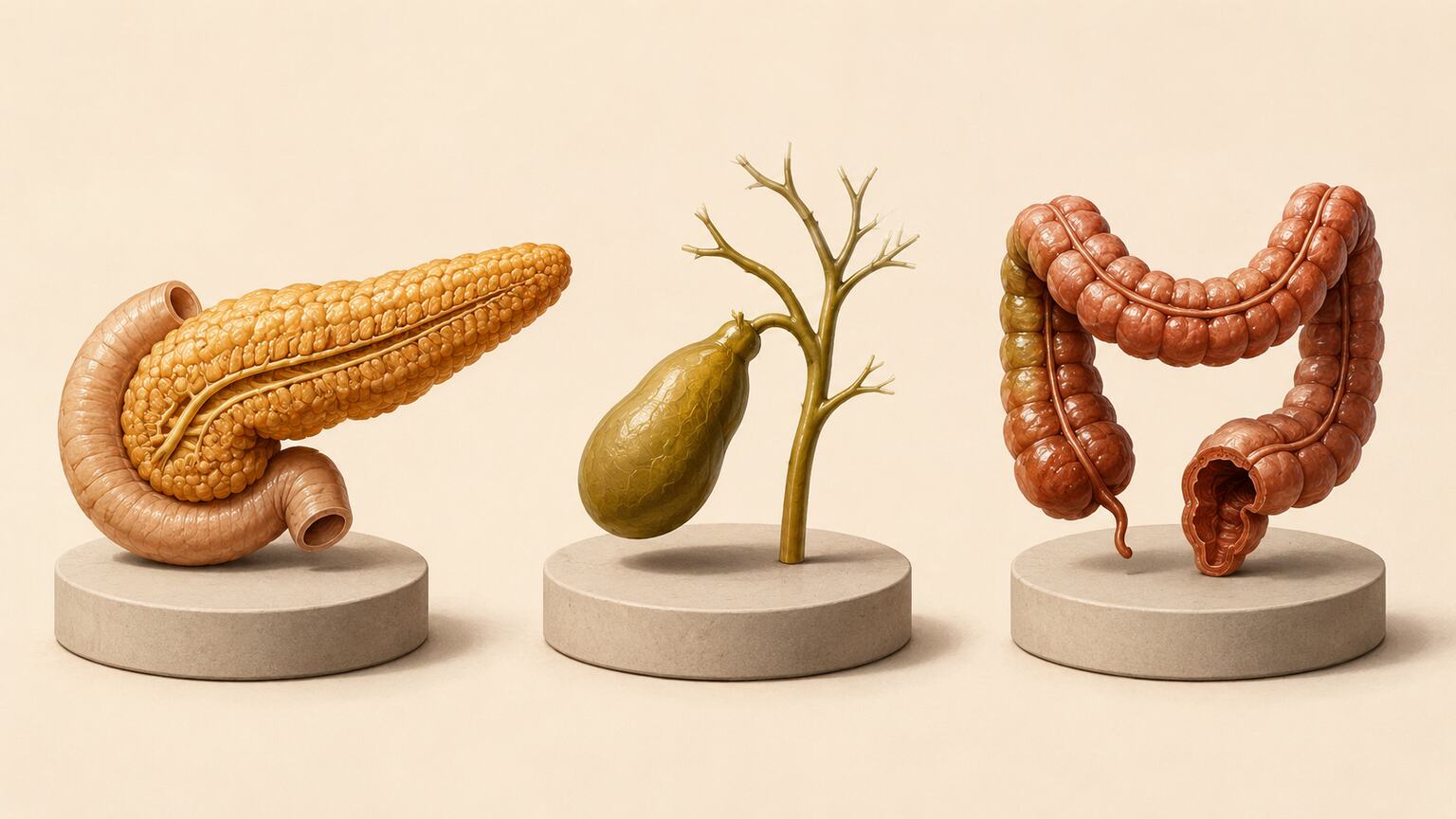
Everything covered so far treats the magnitude scale as if it applies identically everywhere, but the specific numeric cutoffs actually shift depending on which cancer is being evaluated, since each organ system produces and clears CA 19-9 somewhat differently and has been studied separately. Ampullary cancer — a rarer cancer of the point where the bile and pancreatic ducts join the small intestine — has its own validated optimal cutoff of 46 U/mL, noticeably lower than the general 37 U/mL baseline most people encounter. Cholangiocarcinoma, cancer of the bile duct itself, uses a cutoff of 103 U/L, above which studies have found a significant association with metastasis and reduced survival at three, six, and twelve months. Colorectal cancer with liver metastasis has its own separately established cutoff of roughly 35.24 U/mL, distinct from the pancreatic-focused numbers most discussions of this marker default to.
The practical implication is that a result of, say, 60 U/mL means something different depending on which cancer, if any, is actually being investigated. In the context of suspected ampullary or bile duct cancer, that number has already crossed a clinically meaningful line; in the context of general pancreatic cancer screening, it's barely past the starting point covered earlier in this article. This is exactly why the specific clinical context your doctor is working within matters as much as the raw number itself, and why the same CA 19-9 result can prompt very different levels of concern from two different specialists evaluating two different possible diagnoses.
None of these disease-specific cutoffs were pulled from thin air, either — each was derived by researchers testing a range of possible dividing lines against confirmed outcomes in real patient cohorts and selecting whichever value best balanced catching true cases against avoiding false alarms for that specific cancer. That's worth knowing simply because it explains why these numbers look so oddly specific rather than being tidy round figures like 50 or 100 — they're the output of real statistical optimization, not an arbitrary convention.
Above 1,000 U/mL: High Specificity, But Still Not Proof
Once CA 19-9 climbs past 1,000 U/mL, its specificity for a genuine digestive-system cancer rises above 99% — meaning a result this high is overwhelmingly more likely to reflect real, significant disease than a borderline number just over 37. Doctors take a result in this range seriously, and appropriately so. But "overwhelmingly more likely" still isn't "certain," and as our companion article on benign causes of an elevated CA 19-9 covers, documented false positives from pancreatitis, cirrhosis, and bile duct obstruction do still occur even above this threshold, which is exactly why imaging and clinical context remain non-negotiable at every level of this scale, no matter how high the number climbs.
There's an important trade-off worth understanding at this exact point on the scale, because it cuts against the intuition that "higher is always more informative." At the 1,000 U/mL threshold, specificity is excellent, but sensitivity for pancreatic cancer actually drops to around 41%, since a substantial share of real, confirmed pancreatic cancers — particularly smaller or earlier-stage ones — never climb anywhere near this level in the first place. In practical terms: a result above 1,000 is a strong signal when it's present, but its absence tells you comparatively little, since plenty of genuine cancers sit well below this line throughout much of their course.
The Extremes: What Happens at 50,000, 100,000, and Beyond
This is where the numbers stop behaving the way intuition suggests they should. The highest CA 19-9 level ever reported in the medical literature belongs to a patient with adenocarcinoma of the tail of the pancreas with liver metastases: a staggering 19,516,020 U/mL — nearly 20 million, a number so far outside ordinary clinical experience that the case was published specifically because nothing like it had been recorded before. That case fits the general pattern: extremely high numbers do tend to belong to extensive, metastatic malignant disease.
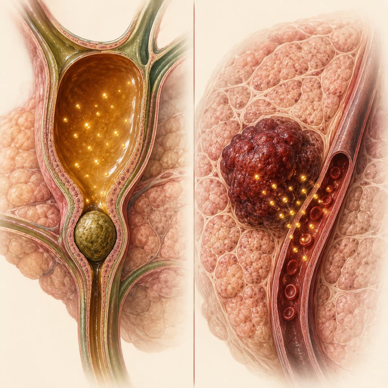
But the pattern breaks down at the very top in a genuinely counterintuitive way. No published case of pancreatic malignancy has ever been recorded above 100,000 U/mL. Meanwhile, a case of simple choledocholithiasis — common bile duct stones, an entirely benign and highly treatable condition — reached 187,000 U/mL, higher than any documented cancer case under six figures. A separate case report describes a benign common bile duct stone driving CA 19-9 to 60,000 U/mL. The mechanism, as covered in more depth in our related article on benign causes, is straightforward once you know it: a completely obstructed bile duct gives CA 19-9 nowhere to go, and pure mechanical blockage can, in rare cases, outpace even the most biologically aggressive tumor's raw production rate. It's a genuinely humbling data point for anyone tempted to treat "the number" as the whole story — even at the far, seemingly unambiguous end of the scale, magnitude alone was not enough to correctly sort these two specific cases.
What separated these extreme cases in practice wasn't the CA 19-9 number at all — it was imaging. In each of the benign case reports described above, a scan showed exactly what was actually happening: a visibly dilated, obstructed bile duct with a stone lodged inside it, not a mass. Once the obstruction was identified and treated, whether through endoscopic removal of the stone or another intervention to restore bile flow, CA 19-9 fell dramatically in every documented case, often back into the normal range within weeks. That fast, clean resolution is itself informative — a malignant process generally doesn't reverse itself nearly that quickly or completely once appropriate treatment for the presumed benign cause begins, which gives doctors a second, time-based way to distinguish the two possibilities beyond the number and the initial scan alone.
Why Doesn't the Number Scale With Tumor Size the Way You'd Expect
It's tempting to assume a bigger tumor automatically makes a bigger number, but research has specifically found that CA 19-9 correlates poorly with tumor size. Instead, the level appears to reflect the tumor's underlying biology — how differentiated its cells are, how actively it's expressing the enzymes needed to build and shed the antigen — more than how many cubic centimeters it occupies. A smaller, more aggressive or poorly differentiated tumor can sometimes produce a disproportionately high number, while a larger, slower-growing one produces comparatively little. This is part of why doctors have never treated CA 19-9 as a stand-in for tumor size on imaging, and why a scan and a blood draw are read together rather than either one substituting for the other.
This decoupling between size and secretion actually makes CA 19-9 more clinically useful in one specific way, not less: it means the marker is capturing something a ruler or a volume measurement on a CT scan genuinely cannot see on its own — a signal closer to how biologically active or aggressive the tumor's cells are, rather than simply how much space they occupy. Two tumors of identical size can carry meaningfully different outlooks if one is producing far more CA 19-9 than the other, which is exactly the kind of complementary information imaging and blood work are meant to provide together rather than either one duplicating the other.
How Doctors Actually Use Magnitude in a Real Decision
In practice, oncologists and surgeons rarely look at a CA 19-9 value as a number in isolation, even once they know roughly which range it falls into. It gets weighed alongside imaging findings, sometimes alongside staging laparoscopy — a minimally invasive surgical look inside the abdomen specifically to check for spread that scans might miss — and always alongside the trend across repeat draws rather than a single moment in time. A very high number that lines up with a large mass and visible spread on a CT scan carries far more weight than the same number in someone whose imaging looks comparatively clean, and a magnitude discussion at any threshold in this article is really shorthand for "how strongly should this result shift our suspicion," not a final verdict delivered by the lab alone.
Research specifically looking at staging laparoscopy has found that a patient's CA 19-9 level going into that procedure meaningfully predicts what the surgical team is likely to find once they actually look inside — higher preoperative levels correlating with a higher chance of discovering previously unseen spread that changes the surgical plan on the spot. That's a good concrete example of magnitude being used exactly as intended: not as a standalone diagnosis, but as one input that helps a care team decide how much additional investigation a specific case warrants before committing to a major operation.
What a High Number Doesn't Tell You

Magnitude has real limits worth naming directly. Roughly 5% to 10% of people are Lewis-negative and cannot produce CA 19-9 at all, meaning a low or normal number in this group carries none of the reassurance it would in someone with normal Lewis status — magnitude simply doesn't apply to them in the way this article describes. On the opposite side, obstructive jaundice from benign causes has been documented to produce false-positive elevations in an estimated 10% to 60% of cases depending on the population studied, a wide enough range that jaundice alone should prompt real caution before treating any number, however high, as conclusive. Both limitations exist at every point on the magnitude scale, not just at the extremes.
These two limitations pull in opposite directions, which is itself worth appreciating. Lewis-negative status means the scale described throughout this article can understate a real problem, keeping a genuinely serious situation looking falsely reassuring. Jaundice-driven false positives mean the same scale can overstate a problem, making a benign situation look falsely alarming. A single magnitude reading, without knowing which of these two distortions might be in play for a specific patient, is genuinely incomplete information on its own — which is exactly why a thorough workup always asks about both possibilities rather than assuming the number is speaking for itself.
Age and general health status add a third, subtler layer worth mentioning. Older patients and those with multiple coexisting chronic conditions are more likely to have at least one of the benign contributors described throughout this article's companion piece on benign causes already present in the background, which can shift a given magnitude reading upward slightly even before any new disease process is considered. None of this means magnitude is unreliable or not worth paying attention to — it means it's one genuinely informative piece of a larger puzzle, read most accurately by someone who also knows the rest of a patient's health picture.
What to Ask Your Doctor When Your Number Is High
A specific, magnitude-aware question tends to get a more useful answer than a general one. It's worth asking directly which range your result falls into by the standards your specific care team uses, since cutoffs vary somewhat between institutions and cancer types. Ask whether imaging has been done or is planned, since a number at any level covered in this article means comparatively little without a scan to correlate it against. And if your result is genuinely extreme — in the tens of thousands or higher — it's entirely reasonable to ask directly whether a benign, mechanical cause like a bile duct obstruction has been considered and ruled out before assuming the number reflects malignancy, given how clearly the case reports above demonstrate that possibility.
It's also worth asking what would need to happen for your care team to feel confident either way — a specific imaging finding, a specific repeat value, a specific procedure — rather than leaving the timeline open-ended. Given how much this article's own case reports demonstrate about how quickly a benign, mechanical cause can resolve once identified and treated, knowing the concrete next milestone your team is watching for is often more reassuring than the raw number itself, however high or low it happens to be on the day you first see it.
Frequently Asked Questions
Does a higher CA 19-9 number always mean a worse prognosis?
Generally, higher levels correlate with more advanced or metastatic disease when cancer is confirmed, but the relationship isn't perfectly linear, and extremely high numbers have also been documented in entirely benign conditions like bile duct obstruction.
What CA 19-9 level is considered a cutoff for unresectable pancreatic cancer?
Different studies have proposed different cutoffs, ranging from around 150 U/mL (88% positive predictive value for unresectability) to 500 U/mL, which an international consensus uses as one criterion for classifying a tumor as borderline resectable.
Can a benign condition really cause a higher CA 19-9 than cancer?
Yes, in documented cases at the extreme end of the scale. A benign bile duct stone case reached 187,000 U/mL, higher than any published pancreatic cancer case below 100,000 U/mL, because complete bile duct obstruction can outpace even aggressive tumor production.
Does CA 19-9 level correlate with how big the tumor is?
Not reliably. Research has found CA 19-9 correlates poorly with tumor size, since the level appears to reflect the tumor's underlying biological activity more than its physical volume.
Is there a CA 19-9 level so high it's automatically cancer?
No single number is automatic proof. Even at 60,000 to 187,000 U/mL, documented benign causes exist, which is why imaging and clinical context remain necessary at every level of this scale.
What should I do if my CA 19-9 result is very high?
Ask your doctor what range it falls into by their standards, whether imaging has confirmed or ruled out a mass or obstruction, and what specific next step the number itself is expected to change.
Do different cancers use different CA 19-9 cutoffs?
Yes. Ampullary cancer uses an optimal cutoff of 46 U/mL, cholangiocarcinoma uses 103 U/L, and colorectal cancer with liver metastasis uses roughly 35.24 U/mL, each distinct from the general 37 U/mL figure most commonly cited for pancreatic cancer.
How quickly can a very high CA 19-9 from a benign cause return to normal?
In documented case reports of benign bile duct obstruction, CA 19-9 fell dramatically, often back into the normal range within weeks, once the obstruction was identified and treated, which is itself a useful clue distinguishing a resolving benign cause from ongoing malignancy.
Conclusion
A CA 19-9 result isn't a single alarm that either goes off or doesn't — it's a number that means genuinely different things depending on exactly where it sits along the full range covered in this article, from a mildly flagged 45 to a staggering seven-figure outlier. Higher numbers do, on average, correlate with more advanced disease and a stronger case for malignancy, and the specific cutoffs covered in this article — 100, 150, 336, 500, 1,000 U/mL, and the cancer-specific variations on each — every one marks a real, research-backed shift in how doctors weigh the result. But even the very top of the scale has produced a genuine surprise: a benign bile duct stone outscoring every recorded cancer case under 100,000 U/mL. Magnitude matters, and it deserves to be understood on its own terms — but it has never been, and was never meant to be, the entire answer on its own.
Still Not Sure What Your Results Mean?
Upload your labs and get a complete, visual, plain-language interpretation of every biomarker — delivered to your inbox in under 15 minutes.
Get My ReportThis article is for educational purposes only and does not constitute medical advice. Always consult your healthcare provider regarding your specific lab results.