
Can Liver Disease Affect Your CEA Test Results?
Yes — liver disease can raise your CEA result even when there's no tumor anywhere in your body, and it's a far more common reason for an unexplained elevated result than most people realize. The connection isn't really about the liver "producing" more CEA the way a tumor does. It's about plumbing. Your liver is the organ primarily responsible for pulling CEA out of your bloodstream and breaking it down, and when that organ is inflamed, scarred, fatty, or blocked, CEA backs up in your blood the same way traffic backs up when a highway off-ramp closes — not because more cars suddenly appeared, but because fewer of them can get off the road. Research has found this pattern in the majority of people with chronic liver disease, and understanding exactly how and why it happens can turn a frightening, unexplained number into something you can discuss with real context at your next doctor's visit.
This matters for a genuinely large number of people, because liver conditions like fatty liver disease are extremely common in the United States, often silent, and frequently discovered around the same time as an unrelated blood panel that happens to include a CEA test. Knowing that your liver — not a hidden cancer — could be behind a mildly elevated number is often the single most useful piece of context available before that next conversation with your doctor.
What CEA Actually Is, in Plain Terms
CEA stands for carcinoembryonic antigen — a mouthful of a name that simply means it's a protein normally made in large amounts before birth, during fetal development, and then largely switched off afterward, though small amounts continue to be produced throughout adult life by the lining of the digestive tract. It's a glycoprotein, meaning it's a protein with sugar molecules attached to its surface, and those sugar tags turn out to matter enormously for how your body eventually disposes of it, as you'll see shortly. Doctors order a CEA blood test mainly to help monitor people who have already been diagnosed with certain cancers, most commonly colorectal cancer, tracking whether the number rises or falls over time as a clue about how treatment is working or whether the disease might be coming back.
What CEA was never designed to be is a stand-alone screening test for the general population. It lacks the precision to reliably tell healthy people apart from those with early cancer, which is exactly why it isn't used that way in routine checkups. Most healthy adults who don't smoke keep a CEA result somewhere under about 3 to 5 ng/mL, depending on the specific lab's reference range, and people who smoke tend to run a bit higher — sometimes up into the 5 to 10 ng/mL range — simply as a normal consequence of tobacco exposure, not disease. Against that backdrop, a liver-related elevation usually looks like a modest bump above the top of normal, not a dramatic spike, which is an important detail to keep in mind as this article works through exactly how high liver disease alone tends to push the number.
How Your Liver Normally Clears CEA From Your Blood
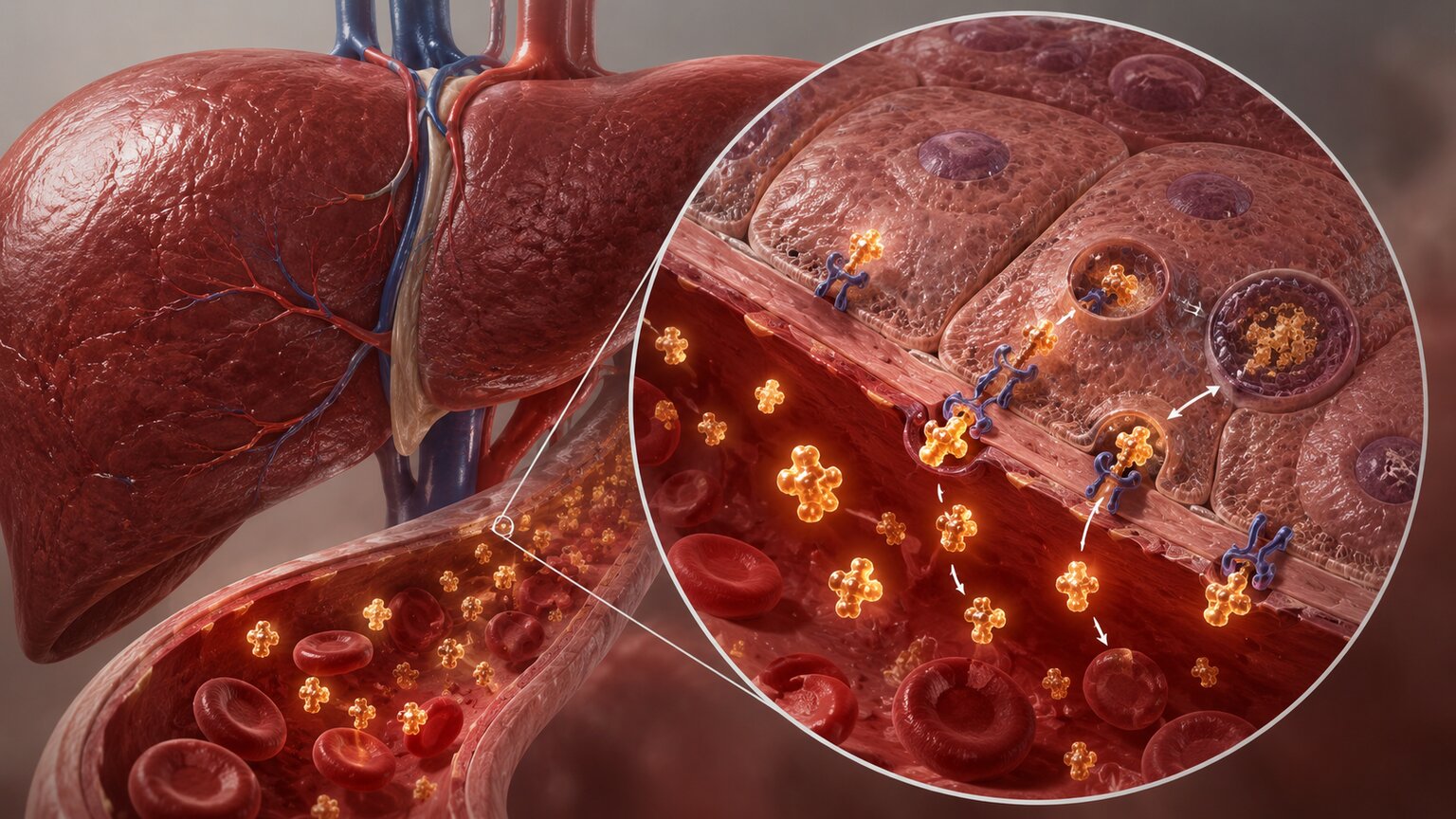
To understand why liver disease throws this system off, it helps to understand how the system works when everything is healthy. CEA doesn't just float around in your bloodstream indefinitely — your body actively removes it, and the liver does most of that work. Research using radioactively labeled CEA found that roughly 70% of it gets cleared from circulation by the liver within just one hour, making the liver by far the dominant organ responsible for keeping CEA levels in check day to day.
The mechanism behind this is a genuinely elegant piece of biology. CEA is a glycoprotein — a protein carrying chains of sugar molecules on its surface, and one of those sugars is called sialic acid, sitting like a cap on the very tip of the chain. When enzymes in your body clip off just a couple of these sialic acid caps, they expose a different sugar underneath called galactose. Specialized structures on the surface of your liver cells, called hepatocytes, act like a lock that specifically recognizes that exposed galactose as a key — this is sometimes referred to as the hepatic binding protein or asialoglycoprotein receptor. Once that recognition happens, the CEA molecule gets pulled inside the liver cell and broken down in a specialized compartment called a lysosome, essentially a tiny recycling center within the cell. Before it even reaches the hepatocyte, research suggests CEA is often first picked up by Kupffer cells — specialized immune cells that live inside the liver and act as an early filtering checkpoint — before being handed off to the hepatocytes for final processing. A smaller portion, around 10% in experimental liver models, gets routed a different way entirely: excreted directly into bile, the digestive fluid your liver produces, rather than broken down internally.
When the Clearance System Breaks Down
Every part of that clearance process depends on a liver that's working normally — healthy hepatocytes with functioning surface receptors, healthy Kupffer cells doing their filtering job, and an open, unobstructed path for bile to flow. Liver disease can interfere with any one of those steps, and when it does, CEA simply doesn't get pulled out of the bloodstream as efficiently as it should. It's not that a diseased liver starts manufacturing new CEA out of nowhere — in most cases, the body's normal, low-level production continues unchanged. What changes is the exit rate, and when less CEA leaves the bloodstream than usual, the concentration that remains in it climbs.
This turns out to be a remarkably common finding once researchers started specifically looking for it. Studies measuring CEA in people with confirmed, biopsy-proven liver disease — with no cancer anywhere in their body — have found the marker elevated in a striking share of cases: roughly 70% of people with chronic liver disease and about 50% of people with acute liver disease showed a falsely elevated CEA driven entirely by their liver condition. Broken down by specific diagnosis, one study found elevated CEA in 71% of people with chronic hepatitis B and 84% of people with chronic hepatitis C. Numbers like these are exactly why an elevated CEA, on its own, is never treated as proof of cancer by anyone who understands how the test actually behaves in the real world.
Curious what your own lab results are really telling you? Upload them and get a complete, plain-language breakdown in under 15 minutes.
Analyze My ResultsCirrhosis: The Condition Studied Most Closely
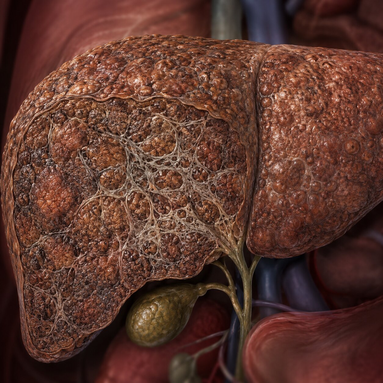
Cirrhosis — permanent scarring of the liver, usually built up over years from causes like chronic alcohol use, hepatitis, or fatty liver disease — has been studied specifically for its effect on CEA, and the findings line up closely with the general clearance story above. One study of cirrhotic patients found that 38.4% had an abnormal CEA level, with no cancer present. Looking more closely at severity using the Child-Pugh classification system, a standard scale doctors use to grade how well or poorly a cirrhotic liver is still functioning, the rate of elevated CEA climbed somewhat as liver function worsened: 28.6% in the mildest category (Child's grade A), 40.6% in the moderate category (grade B), and 42.4% in the most severe category (grade C). That trend toward higher CEA with worse liver function fits neatly with the clearance explanation — the sicker the liver, the less efficiently it can pull CEA out of the blood — though the researchers noted this particular trend didn't reach statistical significance in their specific patient group.
Just as important as the percentage of people affected is how high the number actually climbed. The mean CEA level among the affected cirrhotic patients in that study was 4.75 ng/mL — a real, measurable elevation compared to healthy people, but nowhere close to the kind of number that would typically prompt an urgent cancer workup on its own. The researchers also found a meaningful correlation between CEA and other liver blood tests, including one called glycocholic acid, a bile-acid marker that reflects how well the liver is processing bile — further supporting the idea that this elevation was a liver-function story, not a tumor story. Their stated conclusion was direct: moderate elevations of serum CEA can be expected in cirrhotic patients independent of any malignancy, and clinicians should be cautious about over-interpreting a mildly elevated result in someone already known to have cirrhosis.
Hepatitis and Alcohol-Related Liver Disease
Viral hepatitis follows a similar pattern to cirrhosis, which makes sense given that untreated chronic hepatitis is one of the more common paths that eventually leads to cirrhosis in the first place. Beyond the elevated rates already mentioned for chronic hepatitis B and C, researchers studying alcoholic liver disease specifically have found something worth understanding in more detail: in patients with alcoholic hepatitis and cirrhosis, examination of liver tissue under the microscope revealed marked proliferation of bile ductules — essentially, an overgrowth of the tiny channels that carry bile within the liver — accompanied by prominent CEA staining directly within those cells. Both findings together were associated with elevated CEA in the blood in more than 80% of the cases studied.
That tissue-level detail is genuinely useful context, because it shows the elevation isn't some abstract lab artifact — it corresponds to something real and visible happening inside the liver itself, just not something malignant. The bile ductules proliferating and lighting up with CEA staining reflect the liver's own reparative and inflammatory response to alcohol-related injury, a process entirely separate from cancer even though it produces a similar-looking number on a lab report. This is part of why a doctor who knows a patient has a history of heavy alcohol use, or a confirmed hepatitis diagnosis, will typically interpret a mildly elevated CEA quite differently than they would in someone with an entirely clean liver history.
Fatty Liver Disease: The Quiet, Common Culprit
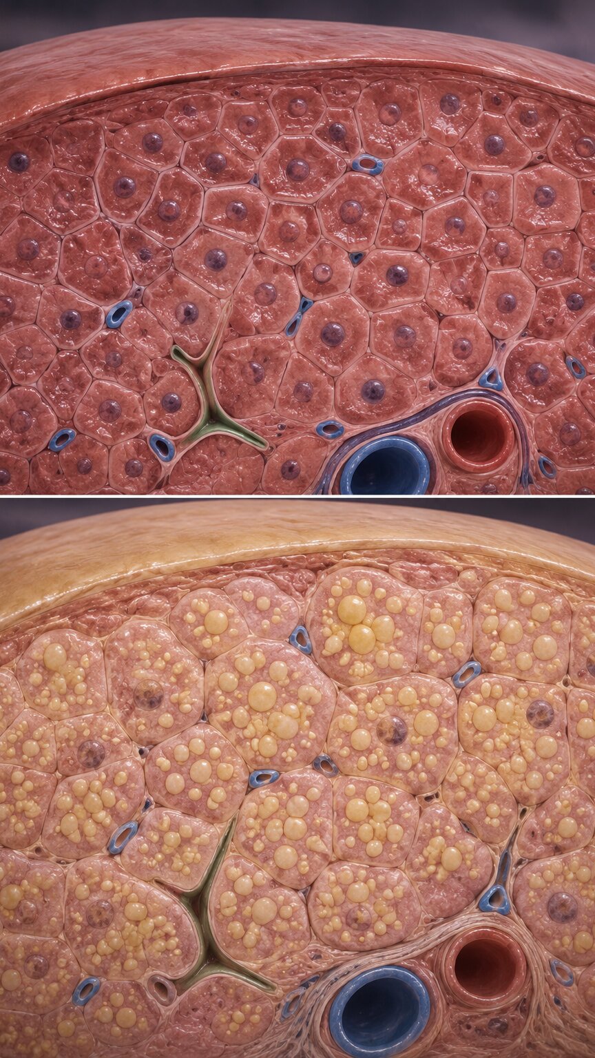
Of every liver condition covered in this article, nonalcoholic fatty liver disease is probably the one most likely to apply to someone reading this without them even knowing it, since it's estimated to affect a substantial share of American adults and frequently causes no symptoms at all. A study of 200 healthy, non-smoking adults in Korea specifically examined whether CEA correlated with fatty liver disease, and found that the prevalence of fatty liver increased significantly as CEA levels rose across the study population, grouped into quartiles from lowest to highest. The average CEA level climbed step by step alongside the grade of hepatic steatosis — the medical term for fat accumulation within liver cells — seen on imaging.
After statistically accounting for other factors that might explain the connection, people in the highest CEA quartile had close to three times the odds of having fatty liver disease compared to those in the lowest quartile, a relationship the researchers described as independent of other metabolic factors. This doesn't necessarily mean falling fatty liver by itself always produces a dramatically elevated CEA in every individual case — the study was looking at population-level patterns, not diagnostic thresholds — but it does add fatty liver disease, arguably the single most common liver condition in the country, to the list of everyday reasons a CEA result might land a little higher than expected in someone who otherwise has nothing else going on.
Bile Duct Blockages and Obstructive Jaundice
Bile is the digestive fluid your liver continuously produces and sends down a system of ducts toward the small intestine, and as mentioned earlier, a portion of your body's CEA is normally excreted directly into that bile as one of its exit routes from the bloodstream. When something physically blocks those ducts — a gallstone lodged in the wrong place, scarring from a condition like primary biliary cirrhosis, or a benign stricture — bile backs up, a condition doctors call cholestasis, and the visible result in more severe cases is obstructive jaundice, the yellowing of the skin and eyes caused by bile pigments building up in the bloodstream instead of draining normally into the gut.
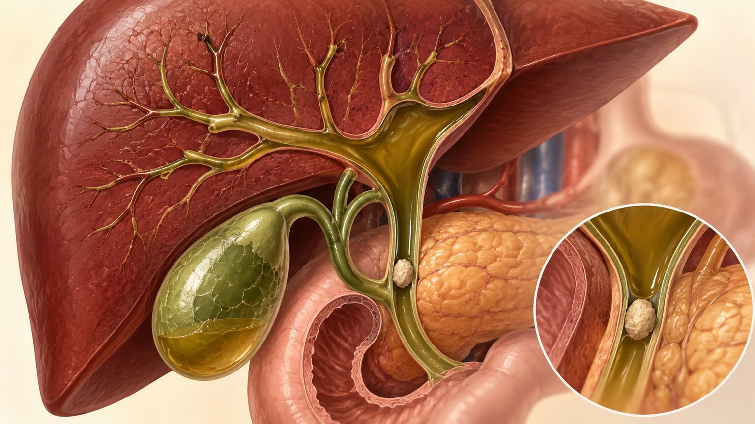
This blockage doesn't just trap bile pigments — research has found that biliary obstruction and cholestasis can independently raise CEA levels, and the proposed explanation is a genuinely interesting one: retained bile appears to have something close to a detergent-like effect on the surface of liver cells, disrupting the very receptors described earlier that are responsible for recognizing and pulling CEA out of the blood in the first place. In other words, a blocked duct doesn't just stop bile from draining — it may actively damage the machinery the liver relies on to clear CEA, compounding the problem from two directions at once. Benign hepatobiliary conditions associated with this kind of elevation include primary biliary cirrhosis, obstructive jaundice from gallstones or strictures, and even hepatic abscess, a localized pocket of infection within the liver. In every one of these cases, the underlying cause is mechanical or infectious, not cancerous, even though the CEA number on the page looks identical either way.
Fulminant Hepatitis: What Rapid Liver Failure Reveals
One especially informative area of research looked at fulminant hepatitis — a rare, severe form of acute liver failure where liver function collapses rapidly, sometimes over just days. In one study following these patients, CEA values ranged from 1.9 to 21 ng/mL, with a mean of 7.0 ng/mL — a real elevation above normal, but still, notably, far lower than levels typically seen in people with actual cancer that has spread to the liver, which this article covers in detail shortly.
What makes this particular study genuinely revealing isn't just the numbers themselves, but their timing. Researchers found that the peak CEA level in these patients occurred later than the point of maximum liver cell death. If CEA were simply leaking out of dying liver cells the way some other liver enzymes do during acute injury, you'd expect the two peaks to line up closely in time. Instead, the delay strongly suggests something different was happening: the rise in CEA wasn't from damaged cells spilling their contents into the blood, but from an already-circulating pool of CEA no longer being cleared properly, because the liver's clearance machinery itself had been knocked offline by the acute injury. That distinction — a traffic jam rather than a spill — is really the theme running through every liver condition covered so far in this article, just expressed in its most extreme and fastest-moving form.
How High Can Liver Disease Alone Push the Number?
This is arguably the single most useful piece of information in this entire article, because it directly answers the question most people actually want answered: how do I know if my elevated CEA is "just my liver" or something more concerning? The research is fairly consistent on this point — benign liver disease, across the different conditions covered above, rarely pushes CEA above about 10 ng/mL. That ceiling holds up remarkably well across cirrhosis (mean 4.75 ng/mL), fulminant hepatitis (mean 7.0 ng/mL, topping out at 21 ng/mL in the most severe case studied), and the general pattern seen across chronic and acute liver disease broadly.

Compare that ceiling to what happens when cancer actually spreads to the liver. One study of colorectal cancer patients found a median CEA of just 6.27 ng/mL in those without liver metastases — already in a similar range to what benign liver disease alone can produce — but a median of 208.1 ng/mL in those whose colorectal cancer had spread to the liver, a roughly thirtyfold difference. Other research has identified a CEA level of about 38.765 ng/mL as a useful cutoff for predicting liver metastases in colorectal cancer, correctly identifying true cases about 91.5% of the time while correctly ruling out cases without metastases about 80.3% of the time in the population studied. A separate line of research has found that CEA values around 30 ng/mL are associated with a higher likelihood that liver lesions can still be surgically removed. Put simply: a CEA of 6 or 8 ng/mL sits comfortably within territory that benign liver disease alone can fully explain, while a CEA climbing into the tens or hundreds tells a genuinely different, much more concerning story that liver dysfunction by itself essentially never produces.
Primary Liver Cancer vs. Cancer That Has Spread to the Liver
There's one more distinction worth understanding, because it involves CEA in a more direct diagnostic role rather than just as a source of confusion. Hepatocellular carcinoma, the most common type of cancer that starts in the liver itself, is primarily monitored using a different marker called AFP (alpha-fetoprotein), not CEA. Research comparing tumor markers across liver cancer types found that AFP and a related marker called PIVKA-II tend to run higher in primary liver cancer — cancer that started in the liver — while CEA tends to run higher specifically in metastatic liver cancer, meaning cancer that originated somewhere else in the body, commonly the colon or rectum, and later spread to the liver.
In one comparative study, CEA showed a positive result in 69.6% of confirmed metastatic liver cancer cases, and performed strongly — with an area under the curve of 0.849, a statistical measure where 1.0 represents perfect accuracy — at distinguishing metastatic liver cancer from ordinary chronic liver disease. When CEA was combined with AFP, AFP-L3, and PIVKA-II together, the combined accuracy for telling primary liver cancer apart from metastatic liver cancer climbed even higher, to an area under the curve of 0.957. The practical takeaway is that CEA genuinely does carry diagnostic weight when liver cancer is already suspected — the trick, as this entire article has emphasized, is knowing which magnitude of elevation reflects that kind of serious underlying process versus which magnitude reflects nothing more than a liver working somewhat less efficiently than it used to.
Why This Matters Especially if You've Had Colorectal Cancer

If you've been treated for colorectal cancer, CEA is likely already a familiar number to you, since standard surveillance guidelines call for checking it roughly every three months for the first two years after treatment, then every six months for up to five years total. Serial CEA monitoring like this can flag a possible cancer recurrence around five months earlier, on average, than it would otherwise be caught through imaging or symptoms alone, which is exactly why the test remains such a valuable part of long-term follow-up care.
This is precisely the situation where knowing about the liver connection becomes genuinely practical rather than just interesting. Someone being monitored after colorectal cancer treatment who also happens to develop fatty liver disease, has a history of hepatitis, or drinks alcohol regularly could see their CEA nudge upward for reasons that have nothing to do with cancer returning — and without this context, that kind of bump could understandably cause real alarm during an already anxious period of ongoing surveillance. The reassuring part is that this exact scenario is precisely why doctors never rely on a single CEA value in isolation: they look at the trend over multiple visits, they correlate it with imaging, and they consider what else might independently explain a modest rise. A CEA that climbs from 3 to 6 ng/mL alongside a new fatty liver diagnosis tells a very different story than one that climbs from 3 to 60 ng/mL with no such explanation in sight.
What Doctors Actually Do When Liver Disease Might Be the Explanation
When a doctor sees a mildly elevated CEA in someone with known or suspected liver disease, the workup generally follows a logical sequence rather than jumping straight to alarm. They'll typically look at standard liver function tests alongside the CEA result — markers like ALT, AST, alkaline phosphatase, and bilirubin — to see whether the pattern fits with active liver dysfunction that could reasonably explain the number on its own. They'll consider the magnitude of the elevation against the benchmarks covered earlier in this article, since a CEA of 8 ng/mL and a CEA of 80 ng/mL prompt very different levels of concern even in someone with confirmed liver disease. And rather than reacting to any single result, they'll typically want to see the trend across more than one blood draw, since a number that holds steady or drifts down over time behaves very differently from one that keeps climbing.
It's also worth knowing that the liver isn't the only organ whose dysfunction can distort a CEA result through impaired clearance rather than true overproduction — kidney failure has been associated with a similar false-elevation pattern, for largely analogous reasons involving reduced clearance of circulating proteins generally. That's a useful piece of context if you have both liver and kidney findings on the same panel, since either one — or both together — can independently account for a modestly elevated CEA without any cancer being involved at all.
Frequently Asked Questions
How high can CEA get from liver disease alone, without any cancer?
Research consistently shows benign liver disease rarely pushes CEA above about 10 ng/mL. Studies of cirrhosis found a mean of 4.75 ng/mL, and even fulminant hepatitis, a severe form of acute liver failure, topped out around 21 ng/mL in the most extreme case recorded.
Why does liver disease raise CEA if there's no tumor?
The liver is responsible for clearing about 70% of circulating CEA from the blood within an hour. When liver disease impairs that clearance process, CEA that's normally produced in small amounts simply isn't removed as efficiently, so it accumulates in the bloodstream.
Which liver conditions are most likely to raise CEA?
Cirrhosis, chronic viral hepatitis B and C, alcoholic liver disease, nonalcoholic fatty liver disease, obstructive jaundice from bile duct blockages, and fulminant hepatitis have all been documented to independently raise CEA in research studies.
Can fatty liver disease alone raise CEA even without symptoms?
Yes. A study of 200 healthy non-smokers found CEA levels rose alongside the grade of fatty liver seen on imaging, with the highest CEA group having nearly three times the odds of having fatty liver disease compared to the lowest group.
How do doctors tell a liver-related CEA elevation apart from cancer that has spread to the liver?
Mainly by magnitude and trend. Colorectal cancer with liver metastases typically produces a median CEA around 208 ng/mL, dramatically higher than the roughly 10 ng/mL ceiling seen in benign liver disease, and doctors also correlate the result with imaging and liver function tests.
I've had colorectal cancer treated. Should a small CEA rise alongside a new fatty liver diagnosis worry me?
Not automatically. This is a well-recognized scenario, and doctors weigh a modest CEA rise very differently when it lines up with a plausible liver explanation and a gentle trend, rather than a sharp, unexplained climb with no other cause identified.
Does CEA help diagnose liver cancer itself?
It plays a supporting role. Primary liver cancer (hepatocellular carcinoma) is mainly tracked with a different marker called AFP, while CEA tends to be more elevated in cancer that has spread to the liver from elsewhere, such as the colon.
What should I ask my doctor if my CEA is high and I also have liver disease?
Ask how your CEA level compares to what's typically seen in benign liver disease versus metastatic disease, whether your liver function tests support liver disease as the explanation, and whether they want to recheck the trend before considering further workup.
Conclusion
CEA was designed to help track cancer, but the number on your lab report is really a snapshot of two competing forces at once: how much CEA your body happens to be producing, and how efficiently your liver is clearing it back out. Liver disease — whether that's cirrhosis, hepatitis, fatty liver, or a blocked bile duct — interferes almost entirely with the second half of that equation, not the first, which is exactly why it can raise CEA without a single cancer cell being involved anywhere in the body. The research is genuinely reassuring on magnitude: benign liver conditions, across nearly every study covered in this article, rarely push CEA above roughly 10 ng/mL, while cancer that has spread to the liver typically produces numbers many times higher. If you have a mildly elevated CEA and a known or suspected liver condition, that combination is a well-documented, thoroughly studied pattern — not a coincidence to panic over, but exactly the kind of context that turns an unfamiliar number into one you can discuss clearly and confidently with your doctor.
Still Not Sure What Your Results Mean?
Upload your labs and get a complete, visual, plain-language interpretation of every biomarker — delivered to your inbox in under 15 minutes.
Get My ReportThis article is for educational purposes only and does not constitute medical advice. Always consult your healthcare provider regarding your specific lab results.