
Understanding What Anti-dsDNA Antibodies Indicate
Anti-dsDNA antibodies are one of the more specifically informative findings that can come back after an initial positive ANA screen, precisely because of what they target: your own double-stranded DNA, the genetic material coiled inside virtually every cell in your body. Unlike a general ANA result, which flags a broad, nonspecific category of self-targeting antibodies and shows up in a wide range of conditions (and even in some healthy people), a positive anti-dsDNA result narrows the picture considerably, since this particular autoantibody is strongly, specifically associated with systemic lupus erythematosus. It's also one of the few autoantibodies that meaningfully rises and falls with actual disease activity, rather than simply staying positive indefinitely once it first appears. This article walks through exactly what anti-dsDNA antibodies are, why they're so much more specific than a general ANA result, how they're actually tested, and why they matter directly for tracking lupus activity and protecting kidney function.

Figure 1. Anti-dsDNA antibodies are shaped to specifically recognize the intact, wound structure of double-stranded DNA — a molecular target distinct enough that this antibody rarely appears outside the specific context of lupus.
Why the Immune System Would Ever Target Its Own DNA
Before getting into the specifics of this particular antibody, it's worth sitting with a genuinely strange fact: your immune system is normally built with extensive safeguards specifically designed to prevent it from ever producing antibodies against your own DNA, or against any of your own tissues more broadly. DNA is about as fundamental to "self" as a molecule can get, present identically in nearly every cell throughout your body, which is exactly why an immune response against it represents such a significant breakdown in one of the immune system's most basic organizing principles: distinguishing self from foreign material.
In lupus, this normal safeguard breaks down through a combination of genetic predisposition and environmental triggers that researchers are still actively working to fully map out. Cells throughout the body die naturally as part of ordinary tissue turnover, a process called apoptosis, and this process normally clears away the resulting cellular debris, DNA included, quickly and quietly, without ever presenting it to the immune system in a way that could provoke a response. In lupus, this clearance process appears to work less efficiently than normal, leaving more DNA and other nuclear material available for a longer period, in a context that increases the odds it eventually gets recognized as a target rather than harmlessly cleared away.
Genetic factors influencing this breakdown have been an active area of research for decades, with numerous specific gene variants identified as contributing modestly to overall lupus risk, though no single gene variant is considered necessary or sufficient to cause the disease on its own. Environmental triggers, including certain infections, ultraviolet light exposure, and hormonal factors, are similarly understood to interact with this underlying genetic susceptibility rather than causing lupus directly in isolation, reflecting the genuinely multifactorial nature of how this specific breakdown in self-tolerance ultimately comes about in any individual patient.
What Double-Stranded DNA Actually Is, and Why the Distinction Matters
To understand why this specific antibody carries so much diagnostic weight, it helps to understand exactly what it's targeting. DNA in its normal, functional state exists as a double helix — two complementary strands wound tightly around each other, held together by chemical bonds between paired bases running the length of the molecule. This double-stranded, wound structure is the DNA form found inside the nucleus of virtually every living cell, where it's normally packaged and protected, largely hidden from routine contact with the immune system.
Anti-dsDNA antibodies are specifically shaped to recognize this intact, double-stranded structure, distinguishing them from a broader, related category of antibodies that instead target single-stranded DNA, a less structurally specific, more commonly encountered target that shows up as a nonspecific finding across a much wider range of conditions. This distinction between targeting the double-stranded versus single-stranded form isn't a minor technicality — it's the exact structural specificity that makes anti-dsDNA testing so much more clinically informative than testing for antibodies against DNA in general.
Single-stranded DNA, the form anti-single-stranded-DNA antibodies target, typically arises when the double helix has been unwound or damaged, exposing individual strands that wouldn't normally be presented this way under healthy conditions. Because this unwound, exposed form can result from a wide range of routine cellular processes, tissue damage, and unrelated inflammatory conditions, antibodies against it turn out to be a much less discriminating marker, showing up incidentally across numerous conditions that have nothing to do with lupus specifically. Antibodies against the fully intact, wound double helix, by contrast, require the immune system to specifically recognize and target DNA in its normal, functional, protected state, a considerably more unusual and specific immunological event that occurs overwhelmingly in the specific context of lupus.
This structural distinction is why laboratories specifically design their testing methods, discussed in more detail later in this article, around confirming genuine double-stranded binding rather than simply detecting any antibody that happens to react with DNA in a broad, unqualified sense. Getting this specificity right is precisely what transforms anti-dsDNA testing from a vague, easily-confounded marker into one of the more genuinely useful, targeted pieces of information in the entire autoimmune workup.
Curious what the rest of your autoimmune panel results are telling you? Upload your results and get a complete, plain-language breakdown in under 15 minutes.
Analyze My ResultsWhy Anti-dsDNA Is So Much More Specific Than a General ANA
A positive ANA result on its own is deliberately built to be a highly sensitive screening test, meaning it's designed to catch essentially every case of several different autoimmune conditions, at the acceptable cost of also flagging a meaningful number of people who don't have any autoimmune disease at all, including a portion of healthy people whose immune systems simply happen to produce low levels of self-targeting antibodies without any disease process behind it. This is exactly why a positive ANA, by itself, is treated as a starting point for further investigation rather than a diagnosis.
Anti-dsDNA antibodies sit at the opposite end of this sensitivity-versus-specificity tradeoff. They're found in a meaningfully smaller share of people with lupus than ANA is (roughly 70% of lupus patients test positive at some point, compared to the near-universal positivity seen with ANA), but when anti-dsDNA is genuinely positive, particularly at a high titer, it's a considerably stronger, more specific signal pointing toward lupus specifically, rather than toward the broader category of autoimmune conditions a positive ANA alone might suggest. This is precisely why anti-dsDNA is one of the specific follow-up tests ordered after an initial positive ANA, rather than being used as a first-line screening test on its own.
This sensitivity-specificity tradeoff is a genuinely fundamental concept in laboratory medicine worth understanding directly, not just in the context of this one antibody. A highly sensitive test, like ANA, is deliberately calibrated to catch nearly every true case of the conditions it's screening for, which necessarily means it will also flag some people who don't actually have those conditions — these are false positives, an accepted tradeoff for making sure genuine cases aren't missed. A highly specific test, like anti-dsDNA, is calibrated in the opposite direction: it may miss some genuine cases (a lupus patient who happens to be anti-dsDNA negative, which does occur in roughly 30% of cases), but a positive result carries much stronger confidence that it reflects the real, targeted condition rather than an unrelated cause. Neither property is inherently better than the other — they serve different, complementary roles at different stages of a diagnostic workup, which is exactly why ANA and anti-dsDNA are used together in sequence rather than as interchangeable alternatives to each other.
It's worth being specific about what "roughly 70%" actually means in practice, since this figure isn't a fixed, universal constant. The reported sensitivity of anti-dsDNA for lupus varies somewhat across different studies and different patient populations, generally ranging from around 50% to 90% depending on the specific testing method used, how active a given patient's disease happens to be at the time of testing, and which specific population was studied. Anti-dsDNA positivity also tends to be more common in patients with kidney involvement specifically, given the direct mechanistic link between this antibody and lupus nephritis covered later in this article, which is part of why the reported prevalence can shift depending on how heavily a particular study population is weighted toward patients with this specific complication.
How Anti-dsDNA Is Actually Tested

Figure 2. The Crithidia luciliae assay uses a single-celled organism whose kinetoplast organelle contains pure, circular double-stranded DNA free of any other cellular proteins — a glowing signal at this specific structure confirms genuine, high-specificity dsDNA binding.
Several genuinely different laboratory methods exist for detecting anti-dsDNA antibodies, and understanding which one produced your result matters, since they differ meaningfully in both sensitivity and specificity. ELISA-based testing, the most common and widely available method, is relatively fast, inexpensive, and highly sensitive, but it's also somewhat more prone to detecting lower-affinity antibody binding that may not carry the same clinical significance as a result confirmed through a more specific method.
The Crithidia luciliae immunofluorescence assay uses a clever biological trick to improve specificity: it exposes a patient's blood sample to Crithidia luciliae, a single-celled organism whose kinetoplast, a specialized organelle, contains pure circular double-stranded DNA entirely free of any other proteins that could otherwise cause the kind of nonspecific binding seen in some ELISA results. If a patient's serum contains genuine anti-dsDNA antibodies, they'll bind specifically to this kinetoplast structure, producing a distinctive glow under fluorescence microscopy that a trained technician can directly visualize and confirm. A related, older method called the Farr assay uses radioactively labeled DNA and specifically detects only high-avidity antibody binding, making it historically valued for its strong correlation with genuine disease activity, though it's used less frequently today than ELISA or the Crithidia method due to the practical challenges of working with radioactive materials.
The concept of antibody avidity, mentioned specifically in the context of the Farr assay, deserves its own brief explanation, since it's a genuinely important factor separate from simply whether an antibody binds its target at all. Avidity refers to the overall strength of the binding interaction between an antibody and its target, reflecting not just whether the interaction happens, but how tightly and stably it holds once it does. High-avidity anti-dsDNA antibodies bind their target more strongly and more consistently than low-avidity ones, and this distinction correlates meaningfully with clinical significance — high-avidity antibodies are more strongly associated with genuine, active disease and are more likely to be the ones forming the kind of persistent, tissue-damaging immune complexes discussed in the kidney-involvement section later in this article. This is part of why a test method specifically capable of distinguishing high-avidity from low-avidity binding, like the Farr assay historically, or the Crithidia assay's requirement for genuinely strong binding to produce a visible glow, tends to correlate more reliably with real disease activity than a method that simply detects any binding regardless of strength.
Because these different testing methods aren't perfectly interchangeable, it's genuinely useful to know which specific method produced your own result, particularly if you're comparing results across different lab visits or different laboratories over time. A titer reported from an ELISA test isn't directly comparable, number for number, to a titer reported from a Crithidia-based test, since the two methods use entirely different measurement scales and detect somewhat different aspects of the antibody response. This is part of why consistency in testing method matters for meaningful longitudinal tracking, discussed in more detail in the next section — switching between different testing methods from visit to visit can introduce genuine noise into what should otherwise be a clean, interpretable trend.
Why Anti-dsDNA Levels Rise and Fall With Disease Activity
One of the most clinically useful properties of anti-dsDNA antibodies, setting them apart from many other autoantibodies, is that their levels genuinely fluctuate in a way that tracks with actual lupus disease activity over time. A general ANA result, once positive, typically remains positive indefinitely regardless of whether a person's lupus is currently flaring or well-controlled, making it far less useful as an ongoing monitoring tool after the initial diagnosis has been established. Anti-dsDNA titers, by contrast, frequently rise in the weeks before a clinical lupus flare becomes symptomatically obvious, and tend to fall again as the flare is brought under control with treatment.
This rise-and-fall pattern is why anti-dsDNA titers are one of the specific laboratory values a rheumatologist tracks over time in someone with an established lupus diagnosis, rather than being a test that's only meaningful at the point of initial diagnosis. A rising titer in someone whose lupus has been stable can be an early warning sign prompting closer monitoring or a proactive treatment adjustment, sometimes before a person even notices new symptoms themselves.
It's worth being clear that this rise-and-fall relationship, while genuinely useful, isn't a perfectly reliable, one-to-one predictor in every single case. Some lupus patients show anti-dsDNA titers that track closely and consistently with their disease activity, making the test genuinely valuable as an early warning system specifically for them. Others show titers that remain relatively stable regardless of how their disease is actually behaving clinically, or that fluctuate somewhat independently of symptoms, meaning the test's usefulness as a monitoring tool can vary meaningfully from one individual patient to another. This is part of why an experienced rheumatologist pays attention to how a specific patient's own anti-dsDNA titer has historically related to their disease activity, using that individual pattern, once established over several visits, as context for interpreting future results rather than assuming every patient's titer behaves identically.
The biological reason behind this titer fluctuation connects directly back to the underlying disease process itself. Periods of increased lupus activity generally involve heightened overall immune system activation, including increased production of the specific immune cells responsible for generating anti-dsDNA antibodies in the first place. As this broader immune activation quiets down, whether through the disease naturally settling or through effective treatment, antibody production correspondingly decreases, and existing antibody levels gradually decline as older antibody molecules are naturally cleared from circulation over time. This means the titer isn't simply an arbitrary number that happens to correlate with disease activity — it's a direct, mechanistic readout of how actively the underlying autoimmune process is currently running.
How Titer Levels Are Actually Reported and Interpreted
Anti-dsDNA results are typically reported either as a specific numeric value with an accompanying reference range, or, particularly for immunofluorescence-based testing, as a titer expressed as a ratio, such as 1:10 or 1:80, representing how many times a sample could be diluted while still producing a detectable positive reaction. A higher number in this ratio format indicates a stronger, more concentrated antibody presence, since the sample remained detectably positive even after being diluted to a greater degree.
Laboratories generally establish their own specific reference ranges and cutoff values for what counts as positive, since these can vary somewhat depending on the exact testing method, reagents, and population used to establish that particular lab's normal range. This is part of why comparing a titer reported by one laboratory directly against a titer reported by a different laboratory, without accounting for potential differences in their specific testing methods and reference ranges, can be misleading — the safest, most reliable comparison is generally made using results from the same laboratory and the same testing method over time, exactly the kind of consistent, longitudinal tracking discussed throughout this article.
The Direct Link to Kidney Involvement: Lupus Nephritis
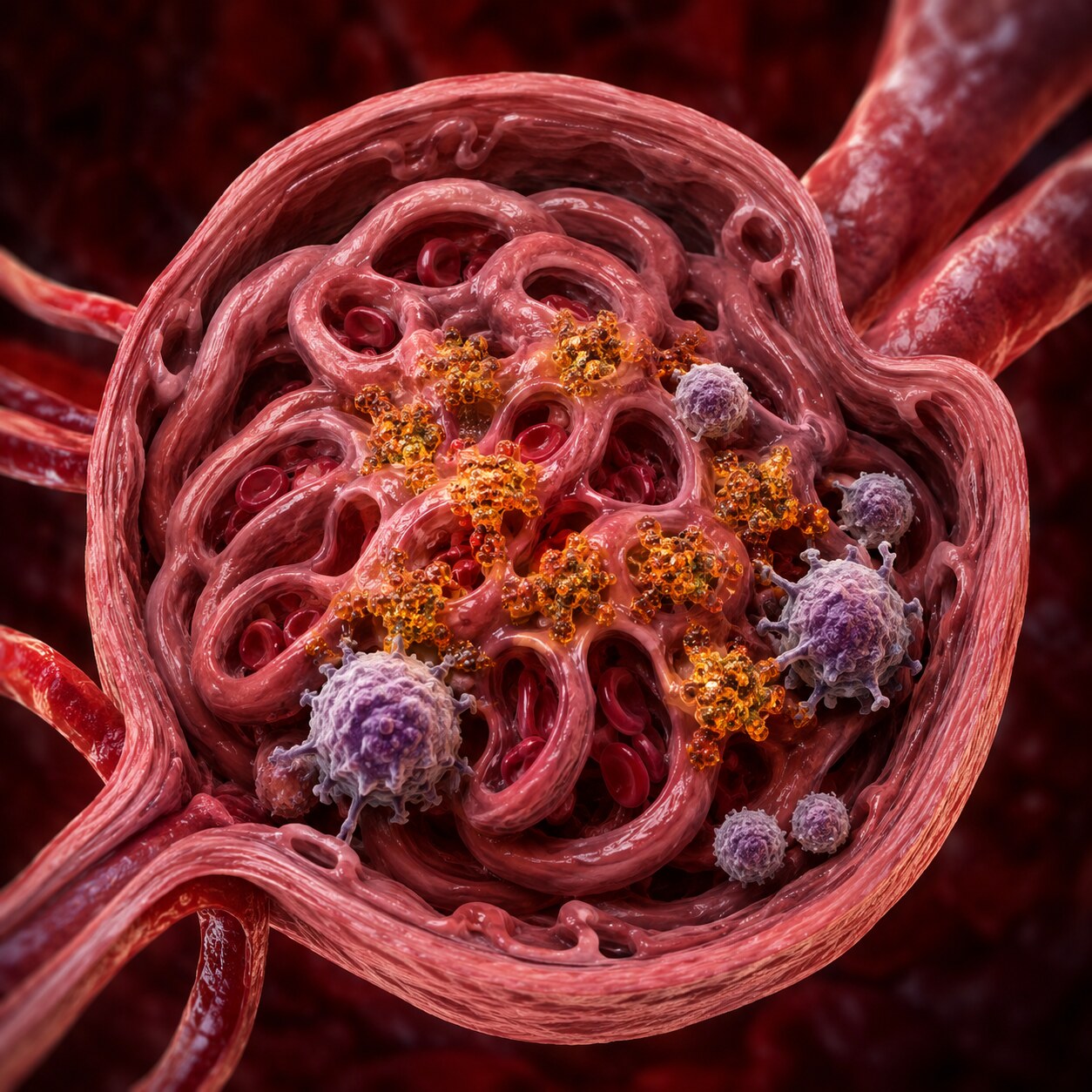
Figure 3. When anti-dsDNA antibodies bind circulating DNA fragments, the resulting immune complexes can become trapped in the kidney's delicate filtering structures, triggering local inflammation — the direct mechanism behind lupus nephritis.
Beyond simply reflecting overall disease activity, anti-dsDNA antibodies play a direct, mechanistic role in one of lupus's most serious potential complications: kidney involvement, called lupus nephritis. When anti-dsDNA antibodies bind to DNA fragments circulating in the bloodstream, they form clumped structures called immune complexes. These complexes can become physically trapped within the kidney's glomeruli, the delicate, tightly woven filtering structures responsible for cleaning your blood, where their presence triggers a local inflammatory response that can progressively damage this filtering tissue if left unaddressed.
This direct mechanistic link is exactly why anti-dsDNA titers are followed particularly closely in lupus patients with any history of kidney involvement, or in those considered at elevated risk for developing it, since a rising titer in this specific context carries additional urgency beyond simply reflecting general disease activity — it's a signal that the same molecular process capable of damaging kidney tissue may currently be more active than usual.
The specific location where these immune complexes deposit within the kidney's filtering structures, and the resulting pattern of inflammation, actually determines which particular subtype of lupus nephritis develops, since kidney specialists classify this condition into several distinct classes based specifically on where and how extensively immune complex deposition and the resulting tissue damage are distributed. This classification, generally confirmed through a kidney biopsy examining the tissue directly, matters considerably for treatment planning, since different classes of lupus nephritis respond differently to different treatment approaches, and some classes carry a meaningfully higher risk of progressing toward more serious, permanent kidney damage than others if left inadequately treated.
Because this classification requires direct tissue examination rather than something a blood test alone can determine, a rising anti-dsDNA titer in someone with signs of possible kidney involvement — protein appearing in the urine, blood in the urine, or a decline in kidney function on standard blood tests — often prompts consideration of a kidney biopsy specifically to characterize which class of lupus nephritis is present and guide the most appropriate treatment approach. The anti-dsDNA result itself doesn't replace this direct tissue evaluation; it serves as one of the key pieces of evidence that helps a clinician decide when pursuing that more invasive, more definitive evaluation is genuinely warranted.
The Complement Connection
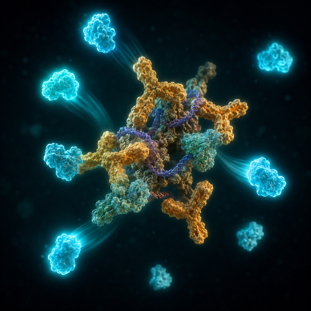
Figure 4. Complement proteins, part of the immune system's normal cleanup machinery, are actively consumed as they bind to and help clear circulating immune complexes — this is why complement levels tend to fall at the same time anti-dsDNA titers rise during active disease.
Anti-dsDNA testing is almost always interpreted alongside a related pair of blood tests measuring complement proteins, specifically C3 and C4, and understanding why requires understanding what complement normally does. Complement proteins are part of the immune system's routine cleanup machinery, helping to identify and clear immune complexes, including the exact antibody-DNA complexes described in the previous section, from circulation before they can cause significant tissue damage.
During a period of high anti-dsDNA activity, larger numbers of immune complexes are being formed, and complement proteins get actively consumed in the process of trying to clear them, causing measured complement levels to drop. This produces a recognizable, complementary pattern that's genuinely useful for tracking lupus activity: a rising anti-dsDNA titer alongside falling C3 and C4 levels is considered a particularly strong combined signal of active, ongoing disease, more informative than either single marker reviewed in isolation, since both are reflecting the same underlying immune complex activity from two different, mutually reinforcing angles.
C3 and C4 are two distinct proteins within the broader complement system, and testing both together, rather than just one, provides a slightly more complete picture than either alone would. The complement system operates through a few genuinely different activation pathways, and C3 sits at a point where multiple of these pathways converge, while C4 is more specifically tied to just one of them. This means C3 and C4 don't always move in perfect lockstep with each other, and a pattern where one drops more than the other can occasionally offer additional clues about which specific complement pathway is most actively engaged in a given patient's disease process, information a rheumatologist can factor into their broader clinical assessment.
It's also worth understanding that low complement levels aren't unique to lupus, just as a positive anti-dsDNA result isn't the only possible explanation for kidney inflammation — several other conditions, including certain other autoimmune diseases and some types of infection, can also consume complement and lower its measured levels. This is exactly why complement testing, like every other individual marker discussed throughout this article, is interpreted as one piece of a larger combined picture rather than a standalone, independently diagnostic result on its own.
When Anti-dsDNA Is Positive, but Symptoms Don't Match
Not every positive anti-dsDNA result reflects active, symptomatic lupus. Some people, particularly those with an established, stable lupus diagnosis, carry a persistently detectable but low-level titer even during periods of genuine clinical remission, without this necessarily signaling an imminent flare. Certain other conditions, and in rare cases healthy people with no autoimmune disease at all, can occasionally show a low-level positive result as well, particularly on the more sensitive ELISA-based testing method described earlier.
This is exactly why a positive anti-dsDNA result, like most laboratory findings covered throughout this article, is interpreted within the full clinical context rather than treated as an automatic, standalone trigger for a treatment change — the specific titer level, the testing method used, the trend over time, and the accompanying complement levels and symptoms together paint a considerably more complete and reliable picture than the single result on its own.
A useful way to think about this mismatch is that the antibody's presence and the disease's clinical activity, while closely linked for most patients most of the time, are ultimately two related but genuinely separate biological phenomena, not two names for the exact same thing. A rheumatologist managing someone with this kind of discordant pattern — antibody present, symptoms and other markers quiet — generally continues routine monitoring rather than intensifying treatment based purely on the antibody level, precisely because treatment decisions in lupus are meant to address active, harmful disease processes, not simply to chase a laboratory number back toward negative for its own sake.
Drug-Induced Lupus: A Different Antibody Pattern
It's worth specifically distinguishing anti-dsDNA-positive lupus from a separate, related condition called drug-induced lupus, since the two are sometimes confused despite involving a genuinely different underlying antibody pattern. Drug-induced lupus, triggered by certain long-term medications, produces lupus-like symptoms but is overwhelmingly associated with a different autoantibody, targeting a nuclear protein complex called histone, rather than double-stranded DNA itself. Anti-dsDNA antibodies are typically negative or only very mildly positive in genuine drug-induced lupus, which is one of the specific clues clinicians use to distinguish this medication-triggered condition from true systemic lupus erythematosus, where anti-dsDNA positivity is a much more central, expected feature.
This distinction carries genuinely practical consequences beyond simple diagnostic labeling. Drug-induced lupus generally resolves once the triggering medication is identified and discontinued, without requiring the kind of longer-term, disease-modifying treatment approach used for true systemic lupus erythematosus. Correctly recognizing the anti-histone-dominant, anti-dsDNA-negative pattern typical of drug-induced lupus can meaningfully shape a treatment plan, potentially avoiding unnecessary long-term immunosuppressive therapy in someone whose condition will resolve simply by stopping the responsible medication, rather than requiring the kind of ongoing management appropriate for genuine systemic lupus erythematosus.
It's also worth noting that a handful of medications have specifically been associated with triggering this drug-induced pattern, and awareness of this connection is part of why a doctor evaluating a new positive autoimmune panel will typically ask specifically about a patient's current and recent medication history, since identifying a plausible triggering medication can meaningfully redirect the entire diagnostic and treatment approach compared to a case where no such trigger is identified.
What Happens After a Confirmed Positive Result

Figure 5. A confirmed positive result is generally followed by a fuller autoimmune workup, including complement levels and a detailed symptom review, rather than being interpreted as a standalone diagnosis based on a single number.
A confirmed positive anti-dsDNA result, particularly alongside a positive ANA and relevant symptoms, typically prompts a fuller evaluation for systemic lupus erythematosus, including the complement testing described earlier, a detailed review of specific lupus-associated symptoms across multiple organ systems, and often additional, more specific autoantibody testing to build a complete diagnostic picture, since lupus diagnosis relies on a defined combination of clinical and laboratory criteria rather than any single test result alone.
This broader evaluation typically also includes a review of basic organ-system screening tests — a urinalysis to check for early signs of kidney involvement, a complete blood count to look for the blood-cell abnormalities lupus can sometimes cause, and a general symptom review covering joints, skin, and other commonly affected systems — since systemic lupus erythematosus is, true to its name, a condition capable of affecting multiple different organ systems simultaneously, and a thorough initial workup is specifically designed to capture that full potential breadth rather than focusing narrowly on the antibody result alone.
For someone with an already-established lupus diagnosis, a rising anti-dsDNA titer, especially alongside falling complement levels, generally prompts closer clinical monitoring and a conversation about whether a treatment adjustment is warranted, potentially catching a developing flare before it progresses to more significant, harder-to-reverse organ involvement.
How frequently anti-dsDNA and complement testing get repeated varies depending on individual disease history and current stability. Someone with well-controlled, quiescent lupus and no history of significant kidney involvement might have these markers checked relatively infrequently, while someone with more active disease, a prior lupus nephritis diagnosis, or a recent flare typically has them monitored considerably more often, sometimes every few weeks during a period of active concern. This individualized monitoring frequency reflects the same underlying principle running throughout this article: the specific clinical context surrounding a given patient shapes not just how a result should be interpreted, but how often it's even worth checking in the first place.

Figure 6. Tracking anti-dsDNA titers and complement levels across multiple visits, rather than reacting to any single result, is what allows a rheumatologist to distinguish a meaningful upward trend from ordinary fluctuation and adjust treatment accordingly.
This longitudinal tracking is part of why lupus follow-up visits so often include a review of previous lab values alongside the current one, rather than treating each new result as an isolated data point. A single anti-dsDNA titer, viewed without its recent history, can be genuinely difficult to interpret with confidence; the same titer viewed as part of a multi-visit trend, moving consistently upward or holding steady, tells a considerably clearer story about what's actually happening, information a single snapshot result could never provide on its own.
Frequently Asked Questions
Does a positive anti-dsDNA result always mean I have lupus?
Not automatically, though it's a strong, specific signal, particularly at a high titer with accompanying symptoms. Lupus diagnosis relies on a combination of clinical and laboratory findings together, not a single test result reviewed in isolation.
Why does my doctor keep rechecking my anti-dsDNA level?
Because, unlike a general ANA result that typically stays positive indefinitely, anti-dsDNA titers genuinely rise and fall with lupus disease activity, making repeated testing a useful way to monitor for a developing flare over time.
Why are C3 and C4 tested alongside anti-dsDNA?
Complement proteins get consumed while helping clear the immune complexes anti-dsDNA antibodies form with circulating DNA. A rising anti-dsDNA titer alongside falling complement levels is a particularly strong combined signal of active disease.
Is a low-level positive result on ELISA as significant as a positive Crithidia result?
Not necessarily. ELISA is more sensitive but can detect lower-affinity binding of less certain clinical significance, while the Crithidia luciliae assay specifically confirms genuine, high-specificity binding to pure double-stranded DNA, generally carrying stronger diagnostic weight.
Can anti-dsDNA antibodies be positive without lupus at all?
It's uncommon but possible, particularly as a low-level result on more sensitive ELISA testing. This is one reason a positive result is generally interpreted alongside symptoms, complement levels, and other findings rather than being treated as diagnostic on its own.
Does a negative anti-dsDNA result rule out lupus?
No. Roughly 30% of people with confirmed lupus never test positive for anti-dsDNA, so a negative result lowers the likelihood but doesn't rule out the diagnosis, especially if other clinical and laboratory criteria for lupus are present.
Conclusion
Anti-dsDNA antibodies target your own double-stranded DNA directly, and that specific molecular target is exactly what makes this antibody so much more clinically informative than a general ANA result — strongly associated with systemic lupus erythematosus specifically, genuinely tracking real disease activity over time, and directly implicated in the immune complex formation that can damage the kidneys through lupus nephritis. Understanding why this antibody rises and falls, why it's tested alongside complement levels, and why the specific testing method matters is exactly what turns a single positive result into genuinely useful, actionable information about what's happening in your immune system right now, not just a historical marker of past disease.
If your own results include a positive anti-dsDNA finding, understanding which of the specific mechanisms and contexts covered in this article most plausibly applies to your situation — a new diagnostic workup, a possible flare in established disease, or a discordant result that doesn't fully match your current symptoms — is exactly the kind of grounded, informed starting point that makes a conversation with your rheumatologist considerably more productive than approaching the number as an unexplained, isolated mystery, and it's a far more useful lens than trying to interpret the titer alone, without any of the surrounding context this article has laid out.
Still Not Sure What Your Results Mean?
Upload your labs and get a complete, visual, plain-language interpretation of every biomarker — delivered to your inbox in under 15 minutes.
Get My ReportThis article is for educational purposes only and does not constitute medical advice. Always consult your healthcare provider regarding your specific lab results.