
What Does Finding Parasite Larvae in a Sample Mean?
Finding larvae in a sample — whether it's stool, sputum, or a scraping from your skin — is a different and generally more urgent finding than seeing parasite eggs alone. Eggs can sit dormant in a sample for weeks without necessarily proving an organism is actively developing inside you right now. Larvae are the opposite: they are a living, moving, immature stage of a worm that has already hatched and is actively growing, migrating, or in some cases even re-infecting the same person without ever leaving the body. That distinction changes how doctors read the result, how quickly they act on it, and in a handful of specific organisms, it can be the single detail that separates a routine deworming prescription from a medical emergency. This guide walks through exactly what a larvae-positive result means, organism by organism, and why the word "larvae" on a lab report deserves more attention than the word "eggs."
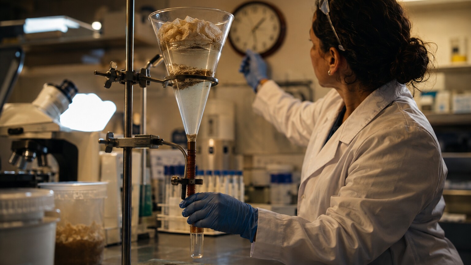
Figure 1. The Baermann funnel technique exploits the fact that live larvae actively migrate toward warmth and moisture, letting them separate themselves from stool debris and settle at the bottom of a collection tube.
It helps to start with what a lab is actually looking at when a report reads "larvae seen" instead of "ova seen" or "cysts identified." A larva is not a piece of a worm, a fragment, or a dead husk left behind after an infection has cleared — it is a self-propelled, developing organism, usually visible moving under the microscope in a fresh, warm sample. Seeing one confirms that a life cycle is actively in progress somewhere in or on the body at the moment the sample was collected, which is a meaningfully stronger statement than what an egg alone can tell a clinician. It also means the timing of the sample collection genuinely matters: a larva seen alive and moving under a microscope reflects what was happening inside the body within hours of that sample being produced, not weeks or months earlier.
Larvae vs. Eggs vs. Adult Worms: Why the Life Stage Matters
Most parasitic worms that infect humans move through several distinct life stages, and a lab report naming the exact stage found is giving you real diagnostic information, not just a technical detail. An egg (also called an ovum) is essentially a sealed capsule — it may or may not still contain a viable, developing organism inside, and depending on the species, an egg passed in stool might need days to weeks of maturation in soil before it becomes capable of infecting anyone at all. A larva, by contrast, has already broken out of that protective shell and is a free-moving, feeding or migrating juvenile worm, which means the biological clock on that particular life cycle is already running.
Adult worms are the final, sexually mature stage, and for many species they never actually appear in a stool sample at all — they stay anchored inside the intestine, shedding eggs or, in some cases, releasing larvae directly. This is precisely why a positive result for larvae is so informative: for the handful of organisms capable of releasing larvae rather than eggs, or capable of completing part of their life cycle inside a single host, finding the larval stage is often the clearest and sometimes the only signal that a mature, egg-or-larvae-producing adult worm is established somewhere in the body right now. In practical terms, this means the same underlying infection can be reported in several different ways depending on which stage a lab happens to catch, and understanding which stage is named on your own report is the first real step toward understanding what it actually means for you.
It's also worth understanding that "larvae" is not a single uniform category the way it might sound. Different parasites release larvae into different parts of the body entirely, which is why the phrase "in a sample" in this guide's title is doing real work — a larvae-positive result might come from a stool sample, but it might just as easily come from a skin scraping, a blood smear drawn at a specific hour of the night, a sputum specimen coughed up during a chest illness, or even a small piece of muscle tissue removed during a biopsy. Each of those sample types corresponds to a different organism's preferred route through the body, and later sections of this guide cover several of the less commonly discussed ones, including a worm whose larvae never leave muscle at all and another whose larvae only circulate in blood at certain hours of the day.
How a Lab Actually Recovers and Identifies Larvae
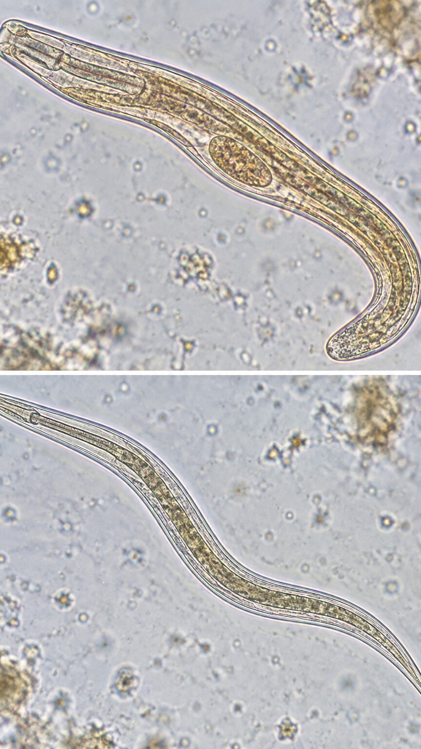
Figure 2. Rhabditiform larvae (left) have a short buccal cavity and a prominent genital primordium, while filariform larvae (right) have a longer esophagus and a notched tail — the two features technicians use to tell them apart under the microscope.
Because larvae are alive and mobile in a fresh sample, labs sometimes use methods built specifically to take advantage of that movement, rather than relying only on a static microscope slide the way they might for eggs or cysts. The Baermann technique, shown above, is a classic example: a stool sample is wrapped in gauze and suspended in warm water inside a funnel, and living larvae, drawn toward the warmth, actively swim or crawl out of the stool and sink to the bottom of a collection tube, where they can be concentrated and examined in far greater numbers than a plain smear would ever catch. A related method, agar plate culture, spreads stool onto a nutrient agar plate and simply waits — larvae crawling across the surface leave visible tracks in the agar as bacteria grow along their trails, sometimes making the infection obvious even before a single larva is directly seen under the scope.
Once larvae are recovered, identifying which species is present comes down to close morphological detail: the length and shape of the esophagus, the presence or absence of a distinct bump called a genital primordium, and the shape of the tail. A rhabditiform larva — short, blunt-tailed, with a wide feeding chamber — usually represents the immature, non-infectious stage still developing inside the gut. A filariform larva — longer, slimmer, with a sharply pointed or notched tail — is typically the mature, infectious stage capable of actively penetrating skin to start a new infection. Telling these two forms apart isn't a minor technicality; for some organisms, it's the single detail that determines whether a finding represents an early, contained infection or one that's already cycling and re-infecting the host.
Sensitivity is the other detail worth understanding before looking at individual organisms, because it directly affects how much weight a single negative result deserves. A single stool sample, examined by a single method, misses a meaningful share of true larval infections — larval shedding can be light, intermittent, or simply timed differently than the day a sample happened to be collected. Studies comparing methods have found that a single Baermann concentration can miss roughly 30% or more of confirmed Strongyloides infections, and combining two or three techniques, or testing on separate days, substantially improves the odds of catching an infection that a single sample would have missed entirely. This is exactly why a doctor who strongly suspects a larval infection based on symptoms or travel history won't necessarily stop looking after one clean result.
Curious what the rest of your lab work is telling you? Upload your results and get a complete, plain-language breakdown in under 15 minutes.
Analyze My ResultsStrongyloides Stercoralis: The Organism Where Larvae Detection Matters Most
If there is one parasite where the word "larvae" on a lab report should immediately get a doctor's full attention, it's Strongyloides stercoralis. Unlike most intestinal worms, Strongyloides doesn't shed eggs into stool at all under normal circumstances — its eggs hatch while still inside the intestinal wall, so what actually appears in a stool sample is the rhabditiform larva, not an egg. This is why stool testing for Strongyloides is fundamentally a search for larvae, and why a lab specifically trained to recognize rhabditiform larval morphology is essential; a technician looking only for eggs, the way they might for a hookworm infection, can miss Strongyloides entirely.
Strongyloides also produces one of the more distinctive skin findings in all of parasitology, called larva currens, Latin for "racing larva." As filariform larvae repeatedly re-penetrate the skin around the anus and buttocks during autoinfection, they leave behind a raised, red, intensely itchy line that can visibly advance several centimeters within just a few hours — dramatically faster than the days-long crawl of cutaneous larva migrans described later in this guide. Because it moves so quickly and often fades within a day only to reappear during a later autoinfection cycle, larva currens is easy to mistake for a simple allergic reaction or hives unless a clinician specifically recognizes the pattern and connects it to a possible Strongyloides infection, particularly in someone with a relevant travel or residence history.
What makes Strongyloides genuinely unusual — and clinically important — is a life cycle feature almost no other human parasite shares: autoinfection. In most worm infections, the larvae that develop inside the host are the non-infectious rhabditiform form, and they need to leave the body, mature in soil, and re-enter through the skin of a new host to keep the cycle going. In Strongyloides, some of the rhabditiform larvae can transform into the infectious filariform stage right there inside the intestine, penetrate the gut wall or the skin around the anus, and re-enter the same person's bloodstream without ever leaving the body at all. That means a Strongyloides infection can persist and quietly renew itself for years, even decades, after someone's last possible exposure — including in people who haven't set foot in a region where the parasite is common since childhood.
Hookworm Larvae: How They Get In and What They Leave Behind
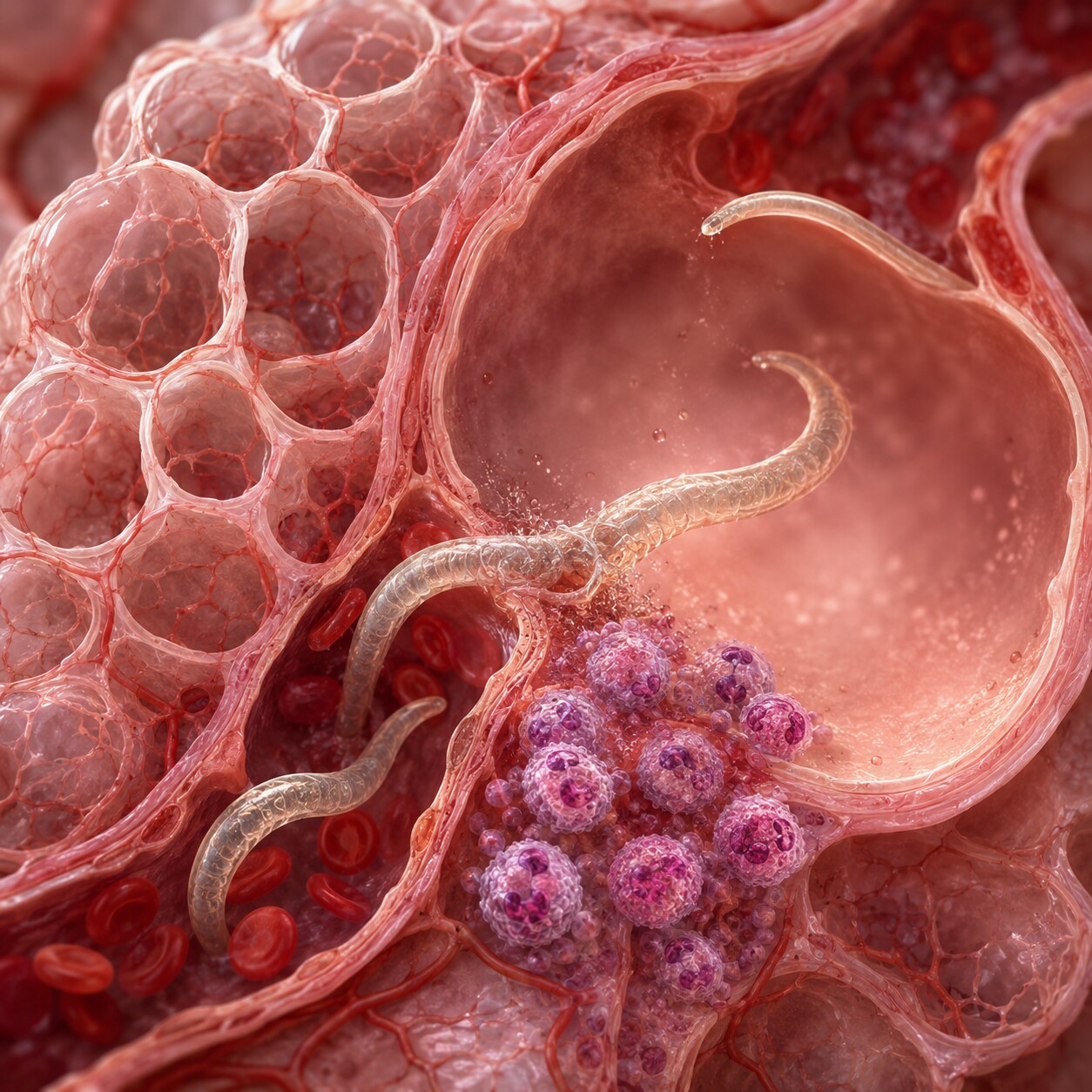
Figure 4. After penetrating skin, hookworm and Ascaris larvae travel through the bloodstream to the lungs, where they break through alveolar capillaries into the airspaces before being coughed up and swallowed back into the gut.
Hookworms — mainly Necator americanus and Ancylostoma duodenale in humans — start their infectious journey as filariform larvae living in warm, moist soil, often where someone has defecated without proper sanitation. These larvae don't need to be swallowed to infect someone; they actively seek out skin contact, most commonly through bare feet, and burrow directly through the skin using enzymes that dissolve their way in within minutes of contact. From there, the larvae enter small blood vessels, travel through the bloodstream to the heart, and are pumped into the lungs, where they break through the tiny air sacs called alveoli, get coughed up the airway, and are swallowed — arriving in the small intestine where they finally mature into adult worms that anchor to the gut lining and begin producing eggs.
That skin-penetration step has a visible signature of its own, and it's the reason a related but distinct condition called cutaneous larva migrans gets its name. When the wrong species of hookworm larva — usually one adapted to dogs or cats, like Ancylostoma braziliense — penetrates human skin, it can't complete its normal migration through a human host and instead wanders aimlessly just beneath the surface of the skin, leaving behind a raised, intensely itchy, winding red track that can advance a few millimeters to a few centimeters per day. It's one of the few parasitic larval infections a person can literally watch happen in real time on their own skin.
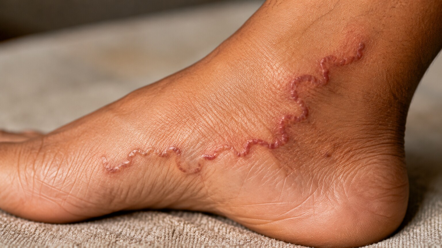
Figure 3. Cutaneous larva migrans occurs when an animal hookworm larva penetrates human skin but cannot complete its normal migration, leaving a visibly advancing track just beneath the surface.
Because hookworm larvae must migrate through the lungs before reaching the gut, an early, heavy exposure can sometimes cause temporary coughing, wheezing, or mild shortness of breath in the one to two weeks after skin penetration, well before any eggs would show up on a stool test. This migratory phase is also where hookworm larvae and Ascaris larvae, despite belonging to very different types of worms, briefly overlap in the kind of respiratory symptoms they can cause — which is covered in more detail next.
Ascaris Larvae and the Lung Phase Called Löffler's Syndrome
Ascaris lumbricoides, the most common intestinal roundworm in humans worldwide, takes a different entry route than hookworm — its eggs are swallowed, usually from contaminated food, water, or soil, rather than penetrating skin directly. But once an Ascaris egg hatches inside the small intestine, the larva that emerges follows a strikingly similar migratory path to hookworm: it burrows through the intestinal wall into the bloodstream, travels to the lungs, breaks into the alveoli, gets coughed up, and is swallowed a second time to finish maturing in the gut. This entire loop, from swallowed egg to fully migrated larva back in the intestine, typically takes about two to three months.
It's during that lung-migration window, roughly one to two weeks after the initial exposure, that a distinctive set of symptoms can appear: a dry cough, wheezing, chest discomfort, and sometimes a low-grade fever, collectively known as Löffler's syndrome. Blood work drawn during this window often shows a striking rise in eosinophils, a type of white blood cell that responds specifically to parasitic and allergic triggers, well before any Ascaris eggs would be detectable in a stool sample — because at that point, the worms are still larvae, migrating through the lungs, and haven't yet reached the intestine to mature and start laying eggs. This is a good example of why the timing of a test, and which stage of the parasite's life cycle it's designed to catch, has to line up with when someone actually got exposed.
Trichinella Spiralis: Larvae That Never Leave Muscle Tissue
Not every larval infection is something a stool sample can ever catch, and Trichinella spiralis is the clearest example why. People become infected by eating undercooked pork or wild game meat — bear, wild boar, and cougar have all been documented sources — that contains microscopic, dormant larvae curled up inside individual muscle fibers. Once swallowed, the larvae are freed by stomach acid, quickly mature into adults in the small intestine, and the resulting new generation of larvae doesn't stay in the gut or migrate through the lungs the way hookworm or Ascaris larvae do. Instead, they burrow directly into the bloodstream and travel specifically to skeletal muscle, where they penetrate individual muscle cells, cause the cell to remodel itself into a protective capsule around them, and settle in for what can be years of dormancy.
Because the larvae end up encysted inside muscle rather than being shed into stool, a standard ova-and-parasite exam is essentially useless for diagnosing trichinellosis — there's nothing circulating in the gut to find. The classic early symptoms instead include swelling around the eyes, muscle pain and tenderness, fever, and a notably high eosinophil count on routine blood work, usually beginning about one to two weeks after eating the contaminated meat, as the newly hatched larvae migrate through the bloodstream toward muscle. Confirming the diagnosis directly requires either a muscle biopsy, where a pathologist can literally see the coiled, encysted larva under a microscope, or more commonly, a blood test looking for antibodies the immune system produces in response to the larvae — since waiting for antibodies to develop takes time, early trichinellosis is often a clinical diagnosis based on symptoms and a food history, treated with albendazole before serology even comes back positive.
Lymphatic Filariasis: Microfilariae Found in a Blood Sample
A different group of worms altogether — the filarial nematodes, spread by mosquito bites rather than contaminated soil or food — release their larval stage directly into the bloodstream instead of the gut, skin, or muscle. In lymphatic filariasis, caused mainly by Wuchereria bancrofti, adult worms live coiled inside the body's lymphatic vessels, the network of channels that normally drains excess fluid back into the bloodstream, and release enormous numbers of a specialized larval stage called microfilariae directly into circulating blood. Unlike the larvae discussed elsewhere in this guide, microfilariae aren't actively migrating toward a new host or a new organ — they're waiting to be picked up by a biting mosquito, which is how the parasite completes its cycle to a new person.
One of the strangest and most clinically important quirks of microfilariae is a pattern called nocturnal periodicity: in most strains of Wuchereria bancrofti, microfilariae are essentially absent from blood drawn during the day and appear in far greater numbers in blood drawn between roughly 10 p.m. and 2 a.m., timed to match the biting habits of the mosquito species that transmits the parasite in a given region. This means a daytime blood draw in someone with a genuine, active infection can come back completely negative for microfilariae, and a lab that isn't specifically told to expect this pattern might report a falsely reassuring result. Diagnosing lymphatic filariasis properly often means either scheduling a blood draw specifically at night, or using a same-day antigen test that detects a protein from the adult worm itself rather than depending on catching circulating larvae at the right hour. Left untreated over years, the chronic inflammation and lymphatic blockage caused by adult worms — not the microfilariae themselves — is what eventually causes the dramatic limb and genital swelling known as elephantiasis, which is why identifying and treating an active infection early matters well beyond the initial lab result.
Why Finding Larvae — Not Just Eggs — Changes the Clinical Picture
Put simply, a larvae-positive result tells a doctor three things an egg-only result usually can't: that the infection is active and developing right now, that a mature, egg-or-larvae-producing adult worm is very likely already established somewhere in the body, and, for the handful of organisms capable of it, that the infection might be capable of renewing itself internally without any further outside exposure. None of that is guesswork read into a name on a report — it follows directly from what a larva biologically is, compared with an egg that may or may not still be viable.
This is also why the specific organism named next to the word "larvae" on a report matters as much as the word itself. A rhabditiform larva identified as Strongyloides carries a very different weight than a filariform hookworm larva recovered from an agar plate culture, even though both are, technically, larvae. Reading a result well means pairing the life stage with the organism's own biology — which is exactly why the sections above walk through each organism separately rather than treating "larvae" as a single, uniform finding.
Geography and personal history add another layer that a lab report alone can't supply, which is why a doctor ordering or interpreting one of these tests will almost always ask about recent travel, where someone has lived over the course of their life, occupational exposure to soil or livestock, and dietary habits like eating undercooked meat or freshwater fish. Hookworm and Strongyloides are most heavily concentrated in tropical and subtropical regions with limited sanitation infrastructure, though Strongyloides in particular has also been documented in pockets of the rural southeastern United States and Appalachia, meaning a domestic history isn't automatically a reason to rule it out. Someone who immigrated decades ago from a region where Strongyloides is common, and who has never had reason to be tested since, is exactly the kind of person a proactive pre-treatment screening protocol exists to protect — long before any larvae would ever show up as an incidental finding on an unrelated test.
This context also explains why a lab technician's first read isn't always the final word. Larval morphology between related species can look similar enough under a routine microscope that a presumptive identification sometimes gets confirmed with additional testing — molecular PCR panels, species-specific antibody tests, or referral to a reference laboratory with more specialized parasitology experience — particularly when the specific species named would change the urgency or drug choice for treatment. A general practice lab flagging "larvae consistent with Strongyloides species" is giving a doctor enough to act on immediately, even before a specialized lab formally confirms the exact identification days later.
Autoinfection and Hyperinfection: The Highest-Stakes Scenario
Figure 6. Corticosteroids and other immunosuppressive drugs can disable the exact immune signals that normally keep Strongyloides autoinfection in check, allowing the larval cycle to accelerate into hyperinfection.
Circling back to Strongyloides, its capacity for autoinfection becomes genuinely dangerous under one specific circumstance: a weakened immune system. In a person with a healthy, fully functioning immune system, the autoinfection cycle tends to stay low-level and contained, sometimes producing no symptoms at all beyond mild, intermittent digestive upset or a recurring, migratory itchy rash where filariform larvae repeatedly re-penetrate the skin near the anus. But certain immune signals, particularly ones involving a group of cells called eosinophils and a signaling pathway type known as Th2 immunity, appear to actively restrain how much the autoinfection cycle can accelerate.
When those restraining signals are suppressed — most commonly by corticosteroid medications, but also by certain chemotherapy regimens, organ transplant immunosuppression, or the virus HTLV-1 — the autoinfection cycle can spiral out of control into a condition called hyperinfection syndrome. In hyperinfection, vastly larger numbers of filariform larvae migrate simultaneously, and they can travel beyond their usual gut-to-lung circuit into the liver, brain, and other organs, sometimes dragging intestinal bacteria along with them into the bloodstream and causing sepsis. Hyperinfection syndrome carries a mortality rate that has been reported as high as 50% or more in some case series, which is precisely why any patient with a known or suspected past Strongyloides infection is typically screened and treated before starting long-term steroids or other immunosuppressive therapy — the goal is to clear the larvae while the immune system can still keep the cycle in check, not after.
Because a stool sample can be falsely negative on any given day even during active hyperinfection, doctors leaning toward this diagnosis in a high-risk patient often don't wait for a positive larvae result before starting treatment — they treat based on risk factors and clinical suspicion alone, then confirm afterward. This is one of the few scenarios in parasitology where serology, a blood test looking for antibodies against Strongyloides rather than the larvae themselves, becomes especially valuable: antibodies can remain detectable even during a stretch when larval shedding happens to be too light for a stool sample to catch, giving clinicians a second, independent way to support the diagnosis when the stakes of missing it are this high.
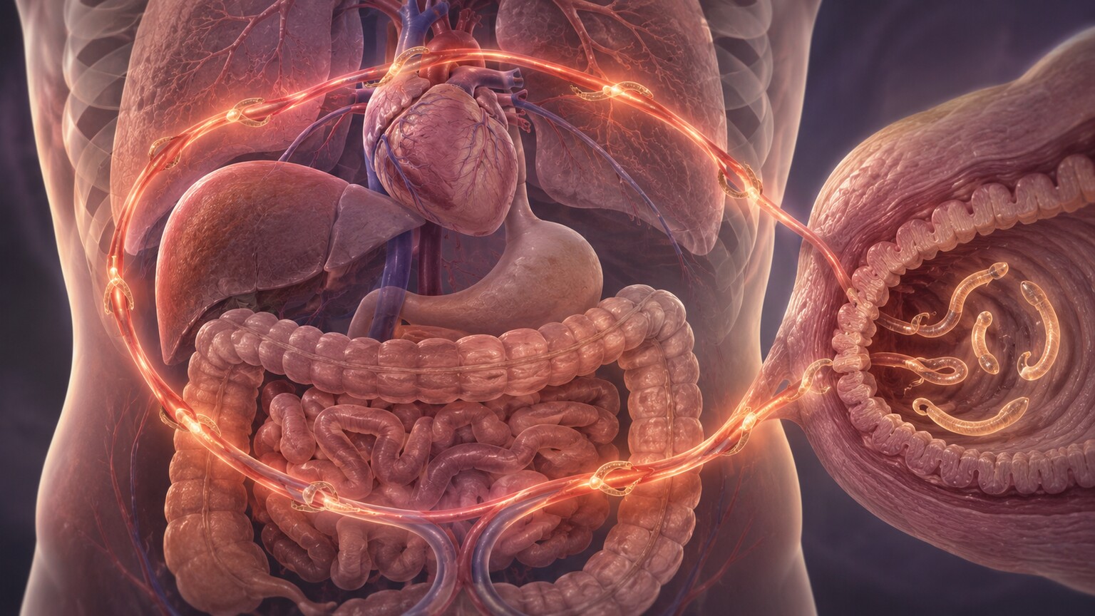
Figure 5. In autoinfection, filariform larvae developing inside the gut re-penetrate the colon wall or perianal skin and re-enter circulation without ever leaving the host, allowing the infection to persist for decades.
What Happens After a Positive Larvae Result
Once larvae are confirmed on a sample, treatment is generally more time-sensitive than it would be for an egg-only finding, precisely because larvae confirm an active, ongoing life cycle rather than a potential or past one. For most hookworm and Ascaris infections, a short course of an antiparasitic medication such as albendazole or mebendazole is typically effective, since these drugs target the adult worms and disrupt the ongoing life cycle at its source. Strongyloides is usually treated differently, most often with ivermectin, which is more effective than the standard benzimidazole drugs specifically because it can reach and kill the migrating larval stages that make this parasite so persistent.
Follow-up testing matters more here than with many other infections, again because of the autoinfection risk carried by Strongyloides in particular. A single negative stool sample after treatment doesn't reliably confirm the infection is fully cleared, since larval shedding can be intermittent even in an ongoing infection — which is why repeat stool testing, sometimes paired with a blood antibody test that can detect past or current exposure even when no larvae happen to be shedding that day, is often used to confirm a true cure before someone is considered fully treated, particularly if they're about to start any medication that suppresses the immune system.
Treatment for organisms whose larvae never circulate in a testable sample, like Trichinella encysted deep in muscle, looks different again. Because the larvae are already walled off inside a protective muscle-cell capsule by the time symptoms typically appear, albendazole is most effective when started early, while newly hatched larvae are still migrating through the bloodstream toward muscle rather than already settled and encysted; anti-inflammatory medication is often added to control the muscle pain and swelling that come from the immune system's reaction to the migrating larvae themselves, which is frequently what actually drives symptoms rather than the parasite doing direct damage. For lymphatic filariasis, a single-dose or short combination antiparasitic regimen can clear circulating microfilariae fairly effectively, though it does less to reverse lymphatic damage that has already occurred from a long-standing adult worm infection, which is one more reason catching an active larval infection early carries real, lasting value.
One practical point applies across nearly every organism covered here: anyone with a known or suspected history of a larva-producing infection, especially Strongyloides, who is about to start corticosteroids, chemotherapy, or any other immune-suppressing treatment should raise that history proactively with their care team before treatment begins, not after symptoms appear. Given how quietly Strongyloides in particular can persist for decades without ever causing a symptom worth mentioning, this is one of the few situations in parasitology where a routine pre-treatment screening test can prevent what would otherwise be a life-threatening emergency.
It's also worth knowing what warrants urgent attention rather than a routine follow-up appointment. Someone with a known Strongyloides history who develops new abdominal pain, persistent vomiting, unexplained fever, confusion, or difficulty breathing — especially soon after starting steroids or another immune-suppressing medication — is describing symptoms consistent with possible hyperinfection, and that combination should prompt an emergency evaluation rather than waiting for a scheduled visit. Outside of that specific high-risk scenario, most larvae findings, including hookworm, Ascaris, Trichinella, and lymphatic filariasis, are manageable on a normal outpatient timeline once identified, with treatment success rates that are generally very high when the right medication is matched to the right organism.
Frequently Asked Questions
Is finding larvae always more serious than finding eggs?
Not automatically more dangerous, but it does confirm an actively developing infection rather than a possible or resolving one. The organism named matters just as much as the life stage — a small number of hookworm larvae recovered by culture is a very different situation from Strongyloides larvae in someone about to start steroids.
Can a Strongyloides infection really last for decades without symptoms?
Yes. Because of autoinfection, Strongyloides can persist at a low, often symptom-free level for 20, 30, or even more than 50 years after the last possible exposure, which is why it's specifically screened for in people with a history of living in or traveling to endemic regions before they start immunosuppressive treatment.
Does one negative stool test rule out larvae after treatment?
Not reliably on its own, especially for Strongyloides. Larval shedding can be intermittent, so doctors typically rely on repeat stool samples over time, sometimes combined with an antibody blood test, before confirming an infection has been fully cleared.
Why do hookworm larvae cause lung symptoms if they infect the gut?
Both hookworm and Ascaris larvae reach the intestine indirectly — after entering the bloodstream, they're carried to the lungs, break into the airways, and are coughed up and swallowed before finishing their development in the gut. That detour through the lungs can cause temporary coughing or wheezing during the migration window.
Why can't a stool test detect Trichinella larvae?
Trichinella larvae leave the intestine almost immediately after hatching and travel to skeletal muscle, where they encyst and stay for years. Since none of the larval stage remains in the gut to be shed, diagnosis instead relies on symptoms, a food history involving undercooked meat, blood antibody testing, or occasionally a muscle biopsy.
Can a daytime blood test miss lymphatic filariasis larvae entirely?
Yes, in many cases. Microfilariae circulating from a Wuchereria bancrofti infection often follow a nocturnal pattern, appearing in far greater numbers in blood drawn late at night than blood drawn during the day. A daytime sample can come back falsely negative unless the lab uses a same-day antigen test instead.
Conclusion
The word "larvae" on a lab report is doing more work than it might first appear to. It tells you that whatever organism is named isn't a leftover trace or a dormant possibility — it's a living, developing stage of a parasite that's actively moving through part of its life cycle inside or on your body right now, whether that sample came from stool, skin, blood, sputum, or muscle. For most organisms, that simply means starting a standard course of antiparasitic treatment and, in some cases, a short round of follow-up testing to confirm the cycle has actually been broken. For Strongyloides specifically, it means understanding a life cycle capable of quietly renewing itself for decades through autoinfection, and taking seriously any conversation with a doctor about immune-suppressing medication before that cycle gets the chance to accelerate into hyperinfection. Either way, a larvae-positive result is worth reading carefully, organism by organism, rather than treating every "positive" the same — the exact species named, the life stage recovered, and the type of sample it came from together tell a far more complete story than the single word "positive" ever could on its own.
Still Not Sure What Your Results Mean?
Upload your labs and get a complete, visual, plain-language interpretation of every biomarker — delivered to your inbox in under 15 minutes.
Get My ReportThis article is for educational purposes only and does not constitute medical advice. Always consult your healthcare provider regarding your specific lab results.