
What Is Considered a Normal Iron Range?
Serum iron — the specific measurement of iron circulating freely in your bloodstream at the exact moment your blood was drawn — has one of the widest normal reference ranges of any routine biomarker, and for good reason: this single number can swing by 30 to 50 percent over the course of a single day in the very same healthy person. Understanding what "normal" actually means for serum iron requires understanding this built-in variability first, since a number that looks alarming at 4 p.m. might be a completely ordinary result if drawn at 8 a.m. instead.

Figure 1. Serum iron follows a natural daily rhythm, typically peaking in the morning and dropping later in the day — a swing large enough that timing alone can shift a healthy person's result meaningfully within the reference range.
The Actual Numbers: Standard Reference Ranges
Most laboratories report a normal serum iron reference range somewhere between approximately 60 and 170 micrograms per deciliter (µg/dL), or roughly 10.7 to 30.4 micromoles per liter (µmol/L) in the alternate SI unit some countries and labs use instead. This is a genuinely wide range compared with many other routine biomarkers, and that width isn't a sign of imprecise testing — it's a direct reflection of how much this specific number naturally fluctuates even in someone with perfectly healthy, well-balanced iron status.
Reference ranges do vary somewhat by sex, since men typically run toward the higher end of this range on average (roughly 65 to 175 µg/dL) while women typically run somewhat lower (roughly 50 to 170 µg/dL), largely reflecting the ongoing iron losses women experience through menstruation. It's worth noting that individual laboratories sometimes report slightly different specific cutoffs depending on their exact testing method and reference population, which is why the specific range printed directly on your own lab report — not a number pulled from a general article — should always be treated as the most authoritative reference point for your own result.
Understanding why reference ranges themselves are established the way they are helps explain why they're deliberately wide rather than narrow. A laboratory reference range is typically built by measuring serum iron in a large sample of presumably healthy individuals and then defining the normal range as the middle 95 percent of those results, meaning roughly 2.5 percent of genuinely healthy people will fall outside the "normal" range purely by statistical definition, on either side, without having any actual iron-related problem at all. This statistical reality is part of why a single value sitting just outside the printed range, especially without any accompanying symptoms or other supporting abnormal findings, isn't automatically treated as clinically significant on its own — it may simply reflect where that particular healthy individual naturally sits within the broader population's normal biological variation.
It's also worth understanding that "micrograms per deciliter" and "micromoles per liter" aren't simply two different-sounding names for the same measurement — they reflect genuinely different unit systems used in different parts of the world, with the conversion factor between them being approximately 0.179 (multiplying µg/dL by this factor converts to µmol/L, and dividing accomplishes the reverse). The United States predominantly uses µg/dL, while many European and other countries predominantly use µmol/L, which is worth knowing if you're comparing a result from a lab report using one unit system against a reference range or article written using the other, since the raw numbers will look completely different despite representing the exact same underlying concentration.
Why Serum Iron Swings So Much More Than Other Biomarkers

Figure 2. Because serum iron peaks in the morning, most providers specifically request a fasting, early-morning blood draw to get the most consistent, comparable reading between different testing occasions.
Unlike ferritin, which reflects your body's overall stored iron reserves and stays relatively stable from day to day, serum iron measures only the small, actively circulating fraction of iron bound to a transport protein called transferrin at that exact moment — and that circulating pool is genuinely dynamic. Iron absorbed from a recent meal, iron being actively shuttled to the bone marrow to build new red blood cells, and iron being released from storage all pass through this same circulating pool constantly, meaning the number reflects a rapidly moving snapshot rather than a stable reserve.
This is precisely why serum iron follows a well-documented diurnal (daily) rhythm: levels typically peak in the morning, often before or shortly after waking, and gradually decline as the day progresses, sometimes dropping by a third or more by evening in the same healthy person. Because of this predictable pattern, most standard testing protocols specifically call for a fasting morning blood draw, both to minimize the effect of a recent meal (particularly one containing iron-rich foods or a supplement) and to capture the level at its most consistent, comparable point in its daily cycle.
The biological mechanism behind this daily rhythm involves a hormone called hepcidin, produced primarily by the liver, which acts as the body's master regulator of iron absorption and release into circulation. Hepcidin levels themselves follow their own daily pattern, generally rising overnight and into the early morning, which suppresses iron release from storage and from dietary absorption during that window, then gradually falling later in the day, which permits more iron to enter circulation. This hepcidin-driven rhythm is layered on top of, and partly explains, the morning-peak pattern seen in serum iron itself, since the overnight period when hepcidin is highest coincides with the period during which the smallest amount of new iron enters circulation relative to what continues being drawn out for use by developing red blood cells in the bone marrow.
Beyond the daily rhythm, serum iron also shows measurable seasonal variation in some populations, with several studies documenting somewhat higher average levels in warmer months compared with colder months, a pattern thought to relate to broader seasonal shifts in diet, physical activity, and possibly vitamin D status, which itself interacts with iron metabolism in ways researchers are still actively working to fully characterize. This seasonal effect is considerably smaller than the daily rhythm and rarely factors into individual clinical decision-making, but it's one more layer of natural variability worth knowing about, particularly for anyone comparing their own results drawn at different times of year.
Menstrual cycle phase introduces yet another layer of natural variation specifically in women of reproductive age, with serum iron tending to run somewhat lower in the days immediately following menstruation (reflecting the blood loss that just occurred) and gradually recovering over the following weeks as the body replenishes what was lost. This is a genuinely normal, expected pattern rather than anything requiring intervention on its own, though it's part of why a single iron panel drawn during or immediately after a particularly heavy period might understate someone's more typical baseline iron status compared with a panel drawn later in the same monthly cycle.
Curious what the rest of your iron panel is really telling you? Upload your results and get a complete, plain-language breakdown in under 15 minutes.
Analyze My ResultsWhy "Normal" Serum Iron Alone Doesn't Rule Out an Iron Problem
Because serum iron is such a moving target, it's genuinely possible for someone with a real, developing iron deficiency to still show a technically normal serum iron reading, particularly early in the process before the body's reserves have been meaningfully depleted. This happens because the body works hard to keep circulating iron levels stable for as long as possible, drawing down stored reserves (reflected in ferritin) well before serum iron itself starts to visibly drop. By the time serum iron has fallen clearly below the normal range, iron deficiency has often already progressed further than a single snapshot number would suggest on its own.
The reverse is also true: a single elevated serum iron reading doesn't automatically confirm iron overload, since a recent iron-rich meal, a multivitamin or iron supplement taken shortly before the blood draw, or even the natural morning peak in someone tested slightly earlier than a comparison sample can all produce a temporarily high number without reflecting any underlying problem. This is exactly why serum iron is almost never interpreted as a standalone result in clinical practice — it's meant to be read alongside total iron-binding capacity (TIBC), transferrin saturation, and ferritin, together forming what's usually called a complete iron panel.
The specific progression from healthy iron status to a fully developed deficiency actually unfolds in three distinct, recognizable stages, and understanding them explains precisely why serum iron lags behind the other markers in showing trouble. Stage one, sometimes called iron depletion, involves declining stored iron reflected by falling ferritin, while serum iron, TIBC, and red blood cell measurements all remain entirely normal, since the body is still successfully compensating using its shrinking reserves. Stage two, iron-deficient erythropoiesis, is where serum iron itself finally starts to fall and TIBC begins rising as the body's reserves become genuinely inadequate to keep circulating iron topped up, though red blood cell size and hemoglobin may still appear normal or only borderline at this point. Stage three, full iron-deficiency anemia, is where hemoglobin and red blood cell size measurements finally drop into clearly abnormal territory, representing the endpoint of a process that, by this stage, has typically been developing for weeks to months already.
This staged progression is precisely why a comprehensive iron workup so often includes a complete blood count alongside the iron panel itself, since red blood cell indices like mean corpuscular volume (MCV) provide yet another independent line of evidence about how far along this process has actually progressed. A patient in stage one, with falling ferritin but everything else still normal, represents a genuinely different clinical urgency and treatment approach than a patient already in stage three with confirmed anemia, even though both might eventually be labeled with the same general "iron deficiency" diagnosis if caught at different points along this same underlying continuum.
How the Full Iron Panel Divides the Diagnostic Work
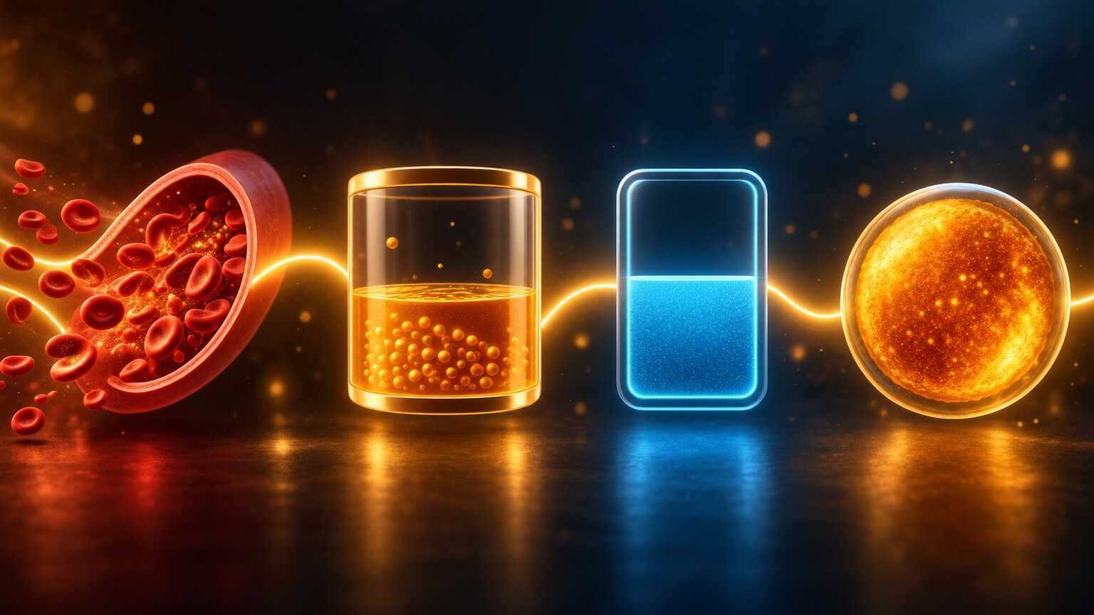
Figure 3. Serum iron is only one of four values that together make up a complete iron panel — each measuring a genuinely different aspect of how your body is handling iron, from moment-to-moment circulation to long-term storage.
Serum iron measures the actual amount of iron circulating in your blood at the moment of the draw. TIBC measures how much additional iron your blood's transport protein, transferrin, could theoretically carry if it were completely full — essentially the total capacity of the transport system, not the amount actually being transported. Transferrin saturation, calculated by dividing serum iron by TIBC, expresses what percentage of that total transport capacity is actually occupied, giving a more stable, ratio-based picture that's somewhat less affected by the moment-to-moment swings of serum iron alone. Ferritin, measured separately, reflects your body's long-term stored iron reserves rather than what's actively circulating, making it the most stable and generally the most sensitive early indicator of developing iron deficiency or overload.
Reading these four values together, rather than any one in isolation, is what allows a provider to distinguish between genuinely different underlying situations that a single serum iron number alone could never separate. Low serum iron with high TIBC and low ferritin points toward true iron deficiency, since the body is actively trying to maximize its iron-carrying capacity to compensate for depleted stores. Low serum iron with low or normal TIBC and normal-to-high ferritin, by contrast, points toward anemia of chronic disease or inflammation, a fundamentally different situation where iron is present in the body but being deliberately withheld from circulation as part of an immune response, requiring a completely different treatment approach.
This distinction matters enormously in practice, since the treatment for these two situations is essentially opposite. True iron deficiency generally responds well to iron supplementation, since the body genuinely needs more iron and will use it appropriately once it's made available. Anemia of chronic disease, by contrast, generally does not respond meaningfully to iron supplementation alone, since the underlying problem isn't a lack of iron in the body but rather the immune system's active decision to withhold available iron from circulation as part of its inflammatory response — giving more iron in this scenario doesn't address the underlying cause and can, in some cases, simply add to iron stores without improving the anemia at all. Correctly distinguishing between these two patterns using the complete panel, rather than assuming based on serum iron alone, directly determines whether a patient receives iron supplements, treatment aimed at the underlying inflammatory or chronic condition, or, in some genuinely mixed cases, a combination of both approaches.
A third, less common but clinically important pattern worth knowing about involves elevated serum iron alongside elevated transferrin saturation and elevated ferritin, which together point toward genuine iron overload rather than deficiency of any kind. This pattern, when it persists consistently across repeat testing rather than reflecting a single transiently elevated reading, is what typically prompts genetic testing for hereditary hemochromatosis or further investigation into other causes of iron overload, since the treatment approach here — therapeutic phlebotomy, essentially controlled bloodletting to reduce total body iron — is entirely different again from either of the two patterns already described.
What Actually Causes Serum Iron to Fall Outside the Normal Range
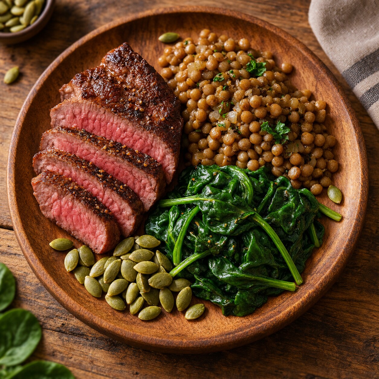
Figure 4. Dietary intake, blood loss, absorption problems, and inflammation are the four broad categories behind most abnormal serum iron results, each requiring a genuinely different follow-up path.
Low serum iron most commonly stems from one of a handful of underlying categories: inadequate dietary intake (particularly in people following strict vegetarian or vegan diets without deliberate attention to iron-rich plant sources), chronic blood loss (heavy menstrual periods, or slow gastrointestinal bleeding from an ulcer, hemorrhoid, or, less commonly, a colon polyp or tumor), impaired absorption from conditions like celiac disease or inflammatory bowel disease, and increased demand during pregnancy or rapid growth periods in childhood and adolescence. Chronic inflammation and certain long-standing infections can also suppress serum iron through a completely different mechanism, one where the body actively sequesters iron away from circulation as a defensive strategy, since many bacteria and other pathogens require iron to grow and multiply.
Elevated serum iron is comparatively less common but has its own distinct set of causes: hereditary hemochromatosis (a genetic condition causing the body to absorb far more dietary iron than it needs), repeated blood transfusions, iron supplement overuse or accidental overdose, certain forms of liver disease, and specific types of anemia in which red blood cell production is impaired despite adequate or excessive iron being available. Distinguishing between these very different possibilities is exactly why an abnormal serum iron result on its own typically prompts the complete panel described above, rather than any specific treatment decision based on serum iron alone.
Hereditary hemochromatosis deserves particular attention among these causes, since it's a genuinely common genetic condition — among the most common inherited disorders in people of Northern European descent — yet frequently goes undiagnosed for years because early symptoms are vague or entirely absent. The condition stems from mutations in a gene called HFE, which normally helps regulate how much dietary iron the intestines absorb; when this regulation fails, the body continues absorbing iron at an inappropriately high rate regardless of how much is already stored, gradually leading to iron accumulation in the liver, heart, joints, and pancreas over years to decades if left untreated. Because symptoms typically don't appear until significant iron accumulation has already occurred, often in a person's forties or fifties, routine iron panel results showing a consistent pattern of elevated serum iron, elevated transferrin saturation, and elevated ferritin — particularly in someone with a family history of the condition — can prompt genetic testing years before symptoms would otherwise have led to a diagnosis, allowing treatment to begin before irreversible organ damage occurs.
Accidental iron overdose deserves a separate, specific mention, particularly relevant to households with young children, since iron-containing supplements and prenatal vitamins are among the most common causes of accidental pediatric poisoning deaths in some regions, precisely because children's iron tablets are often sugar-coated and visually resemble candy. A serum iron level is one of the key tests used in the emergency evaluation of a suspected iron overdose, since the specific level, interpreted alongside the timing since ingestion, directly guides whether specific decontamination or chelation treatment is medically necessary.
Reference Range Differences Across Age and Life Stage
Normal serum iron ranges aren't identical across every stage of life. Newborns and infants naturally run somewhat lower than adult reference ranges during the first months of life, reflecting normal developmental iron physiology rather than any deficiency. Children's ranges gradually shift toward adult values through adolescence, with some variation tied to growth spurts and, in girls, the onset of menstruation. Pregnancy introduces its own distinct pattern: serum iron and ferritin both commonly decline as pregnancy progresses, partly due to the significant iron demands of a growing fetus and expanding maternal blood volume, which is precisely why obstetric care routinely includes iron panel monitoring and often proactive supplementation throughout pregnancy, using pregnancy-adjusted reference ranges rather than standard adult ones.
Infants born full-term generally arrive with sufficient iron stores, largely transferred from the mother during the third trimester of pregnancy, to meet their needs for roughly the first four to six months of life even on an iron-poor diet like breast milk alone, which naturally contains relatively little iron. This built-in reserve is precisely why iron-rich complementary foods, or iron-fortified infant formula and cereals, become specifically important starting around this age window, and why premature infants — who miss out on a meaningful portion of that crucial third-trimester iron transfer — are at meaningfully higher risk of developing iron deficiency earlier and are frequently monitored and supplemented more proactively as a standard part of preterm infant care.
Older adults represent another life stage worth specific mention, since iron deficiency in this population carries a somewhat different differential diagnosis than it does in younger adults. While dietary insufficiency and absorption problems remain relevant, new-onset iron deficiency in an older adult — particularly a postmenopausal woman or any man of any age, since neither group has an obvious ongoing source of blood loss through menstruation — more frequently prompts investigation specifically for occult gastrointestinal bleeding, since colorectal cancer and other significant gastrointestinal pathology become meaningfully more common with advancing age and can present with iron deficiency as an early, sometimes isolated, warning sign well before other symptoms develop.
Medications and Testing Conditions That Can Skew a Single Result
Beyond the natural daily rhythm already covered, several other factors can meaningfully shift a serum iron reading independent of true underlying iron status. Oral contraceptives and hormone replacement therapy have been documented to raise serum iron somewhat in some patients. Recent alcohol consumption can also transiently elevate levels. Iron supplements, including a standard daily multivitamin containing iron, should generally be paused for at least 24 hours before testing when a provider is specifically trying to capture an unsupplemented baseline value, since even one recent dose can meaningfully skew a same-day or next-day result. Because of all these variables, a single unexpected result — particularly one only mildly outside the normal range — is often simply repeated under more carefully controlled conditions before any further workup begins.
Body position and hydration status at the time of the blood draw can introduce smaller, generally minor additional variation as well, since standing versus lying down for an extended period before collection shifts fluid distribution in the bloodstream slightly, mildly concentrating or diluting essentially everything measured in the sample, serum iron included. This effect is small enough that it rarely factors meaningfully into clinical interpretation on its own, but it's one more example of how many subtle, individually minor variables ultimately combine to produce the wide normal range this specific test carries compared with more tightly regulated biomarkers.
Acute illness and infection introduce another significant, often overlooked source of interference, since serum iron drops as part of the body's normal acute-phase response to inflammation regardless of underlying iron status — essentially the same iron-withholding defense mechanism described earlier in the context of chronic disease, but triggered temporarily by something as ordinary as a cold, flu, or minor infection. This is precisely why providers generally avoid ordering an iron panel during an acute illness whenever the testing can reasonably be postponed, since a result drawn during this window can look artificially low, potentially prompting an unnecessary workup for a deficiency that doesn't actually exist once the acute illness resolves and levels return to their genuine baseline within a couple of weeks.
Recent surgery or significant physical trauma produces a broadly similar temporary suppression of serum iron through the same underlying inflammatory mechanism, meaning postoperative iron panels, if drawn too soon after a procedure, can be genuinely difficult to interpret and are generally best deferred by several weeks whenever the clinical situation allows for that kind of delay. Strenuous exercise, particularly endurance activities like long-distance running, has also been documented to transiently lower serum iron in the hours immediately following intense exertion, related to a temporary spike in hepcidin triggered by the exercise itself, which is one more reason standard testing protocols favor a calm, fasting, morning collection over one taken shortly after unusually vigorous physical activity.
How Serum Iron Is Actually Measured in the Lab

Figure 5. Modern automated analyzers measure serum iron using a colorimetric chemical reaction, releasing iron from its transport protein and reacting it with a reagent that produces a color intensity directly proportional to the amount present.
The standard laboratory method for measuring serum iron relies on a colorimetric chemical reaction: a reagent is added to the blood sample that first releases iron from its bound state on transferrin, then reacts specifically with the freed iron to produce a colored compound, with the intensity of that color measured by the analyzer and directly converted into a concentration value. This method has remained the clinical standard for decades because it's fast, highly automatable, and reliably accurate across the wide range of concentrations a serum iron test needs to cover.
Because this method depends on a clean chemical reaction, certain factors can interfere with an accurate reading independent of a patient's actual iron status. Hemolysis — red blood cells rupturing during or shortly after the blood draw itself — releases iron stored inside those cells directly into the surrounding serum sample, artificially inflating the measured result well above the patient's true circulating level. This is one of several reasons an unexpectedly elevated result in an otherwise asymptomatic patient sometimes prompts a repeat draw before any further workup begins, simply to rule out this kind of laboratory artifact rather than a genuine abnormality.
The collection tube itself matters more for iron testing than for many other routine blood tests, since certain tube types contain anticoagulant additives or trace contaminants that can interfere with the colorimetric reaction, either falsely raising or falsely lowering the measured result. Laboratories follow specific, standardized collection protocols using tubes validated as iron-clean specifically to avoid this contamination risk, and this standardization is part of why iron testing generally requires a dedicated blood draw for this specific purpose rather than always being extractable from a leftover sample originally collected for a different, unrelated test.
Turnaround time for a standard serum iron result is typically same-day to next-day at most laboratories, since the colorimetric method itself is fast and highly automated, with the overall panel's turnaround more often limited by batching schedules and courier logistics at smaller labs than by any inherent slowness in the underlying test method itself. This relatively quick turnaround is one reason an iron panel is often a reasonable, low-friction first step when investigating symptoms like fatigue, rather than a specialized test requiring unusual advance planning or a lengthy wait for results.
Symptoms That Prompt Iron Testing in the First Place

Figure 6. Persistent fatigue, unusual paleness, and cold intolerance are among the most common symptoms prompting a first iron panel, though these same symptoms overlap considerably with several unrelated conditions.
Persistent fatigue disproportionate to sleep and activity level, unusual paleness of the skin or inside the lower eyelids, shortness of breath with ordinary activity, unusual cravings for non-food items like ice (a phenomenon called pica specifically associated with iron deficiency), brittle nails, and hair thinning are among the most common symptoms prompting a provider to order an iron panel in the first place. On the opposite end, joint pain, unexplained fatigue paired with a bronze or grayish skin tone, and a family history of hemochromatosis can prompt testing specifically to look for iron overload rather than deficiency.
It's worth noting that essentially all of these symptoms overlap considerably with numerous other, entirely unrelated conditions — thyroid dysfunction, sleep disorders, and depression can all produce very similar fatigue, for instance — which is exactly why a symptom pattern alone never confirms an iron-related diagnosis on its own. The iron panel exists specifically to test that hypothesis directly rather than relying on symptoms alone, and a normal complete panel meaningfully shifts the diagnostic conversation toward these other possible explanations instead.
Pica, the specific craving for ice, dirt, starch, or other non-food substances, deserves a bit more explanation, since it's one of the more distinctive and diagnostically useful symptoms specifically associated with iron deficiency rather than the many other conditions capable of producing generic fatigue. The exact biological mechanism connecting iron deficiency to these particular cravings isn't fully understood, though several theories involve iron's role in specific neurological pathways related to taste and reward processing, and the craving itself — particularly for ice, a pattern specifically called pagophagia — tends to resolve fairly reliably once iron deficiency is properly corrected through supplementation, which is itself considered a reasonably strong piece of supporting clinical evidence that iron deficiency was genuinely the underlying driver.
Restless legs syndrome represents another symptom worth specifically connecting to iron status, since a meaningful proportion of restless legs cases are linked to low brain iron levels, even in patients whose standard peripheral blood iron panel looks entirely normal, since brain iron status doesn't always track perfectly with blood-measured iron status. This is part of why some patients with restless legs syndrome and a borderline-low, though technically still "normal," ferritin result are still offered a trial of iron supplementation specifically to see whether it improves their symptoms, illustrating one of several situations where the standard reference range doesn't necessarily represent the optimal target for every individual patient's specific clinical situation.
Frequently Asked Questions
Why did my serum iron come back normal even though I have classic anemia symptoms?
This is genuinely common, since the body works to keep circulating serum iron stable for as long as possible even as underlying stores decline. Ferritin, which reflects stored iron, often drops earlier and more reliably signals developing deficiency than serum iron alone does at this stage.
Do I need to fast before a serum iron test?
Most standard protocols recommend a fasting, morning blood draw, both to avoid the effect of a recent meal and to capture the level at its most consistent point in its natural daily rhythm, making results more comparable between different testing occasions over time.
Can taking a multivitamin affect my serum iron result?
Yes, meaningfully so. Even a single recent dose of an iron-containing supplement can temporarily raise serum iron. Providers generally recommend pausing iron-containing supplements for at least 24 hours before testing when an unsupplemented baseline reading is specifically needed.
Is a slightly high serum iron result always a sign of hemochromatosis?
No. A mildly elevated result is far more commonly explained by recent diet, supplement timing, or the natural morning peak than by a genetic iron overload condition, which is typically confirmed only after a consistently elevated transferrin saturation across repeat testing and further specific genetic testing.
Why does my provider want the full iron panel instead of just serum iron?
Serum iron alone can't distinguish between several very different underlying situations, including true iron deficiency, anemia of chronic disease, and iron overload. TIBC, transferrin saturation, and ferritin together provide the context needed to tell these apart, since each one measures a genuinely different aspect of iron handling.
Cost and insurance coverage for a standard iron panel are generally straightforward, since it's a routine, widely available, well-established laboratory test typically covered by essentially all health insurance plans when ordered with an appropriate clinical indication like fatigue evaluation or anemia workup. Out-of-pocket cost for patients paying directly is typically modest, reflecting how common and standardized this specific panel is across virtually every clinical laboratory.
Ultimately, the practical value of understanding serum iron's normal range comes down to recognizing what it can and can't tell you on its own. It's a genuinely useful, fast-moving snapshot of one specific piece of a much larger, interconnected system, and its real diagnostic power only emerges once it's read alongside the other three panel components and, when relevant, a complete blood count — never as an isolated number pulled out of context and judged entirely on its own, regardless of how far above or below the printed reference range it happens to fall.
Conclusion
A "normal" serum iron range is genuinely wide, and understanding why — the natural daily rhythm, the influence of recent meals and supplements, and its role as a fast-moving snapshot rather than a stable reserve — explains why this single number is rarely, if ever, interpreted on its own. Reading serum iron alongside TIBC, transferrin saturation, and ferritin turns four individually incomplete pieces of information into a coherent, genuinely diagnostic picture of how your body is actually managing iron, well beyond what any single value could reveal in isolation — and knowing which specific pattern your own results fall into is what ultimately shapes whether the right next step is supplementation, further investigation, or simply reassurance.
Still Not Sure What Your Results Mean?
Upload your labs and get a complete, visual, plain-language interpretation of every biomarker — delivered to your inbox in under 15 minutes.
Get My ReportThis article is for educational purposes only and does not constitute medical advice. Always consult your healthcare provider regarding your specific lab results.